. 2021 Mar 5;128(5):570-584.
doi: 10.1161/CIRCRESAHA.120.318511. Epub 2021 Jan 27.

Influenza Causes MLKL-Driven Cardiac Proteome Remodeling During Convalescence

Affiliations

Influenza Causes MLKL-Driven Cardiac Proteome Remodeling During Convalescence

Yi-Han Lin et al. Circ Res. .

Abstract

Rationale: Patients with and without cardiovascular diseases have been shown to be at risk of influenza-mediated cardiac complications. Recent clinical reports support the notion of a direct link between laboratory-confirmed influenza virus infections and adverse cardiac events.

Objective: Define the molecular mechanisms underlying influenza virus-induced cardiac pathogenesis after resolution of pulmonary infection and the role of necroptosis in this process.

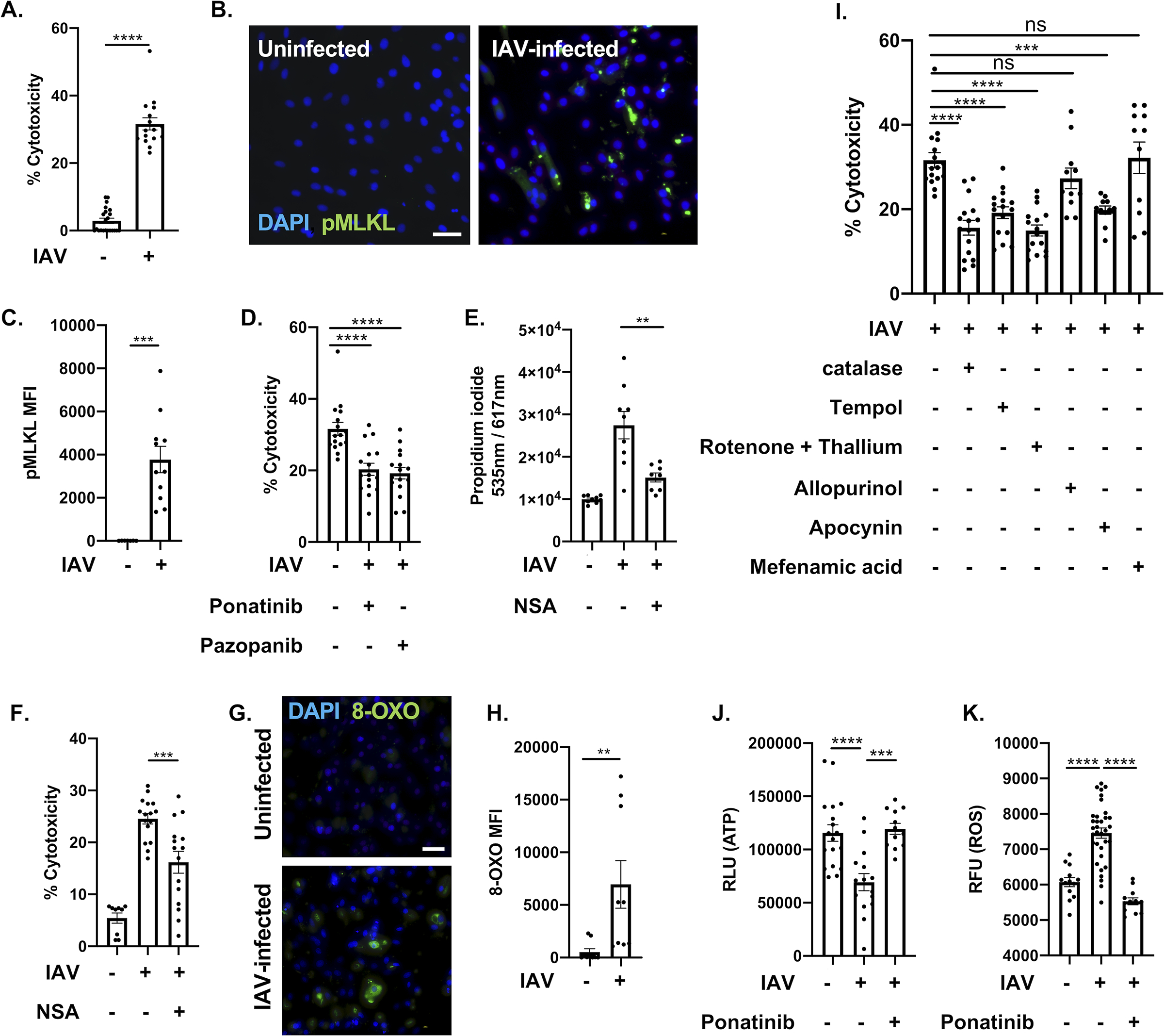
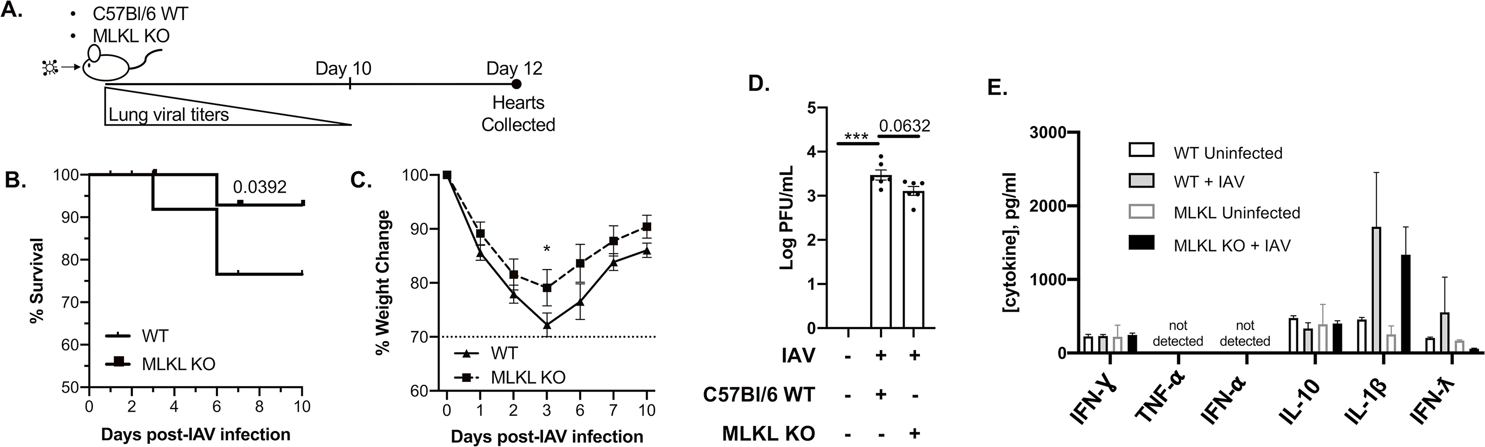
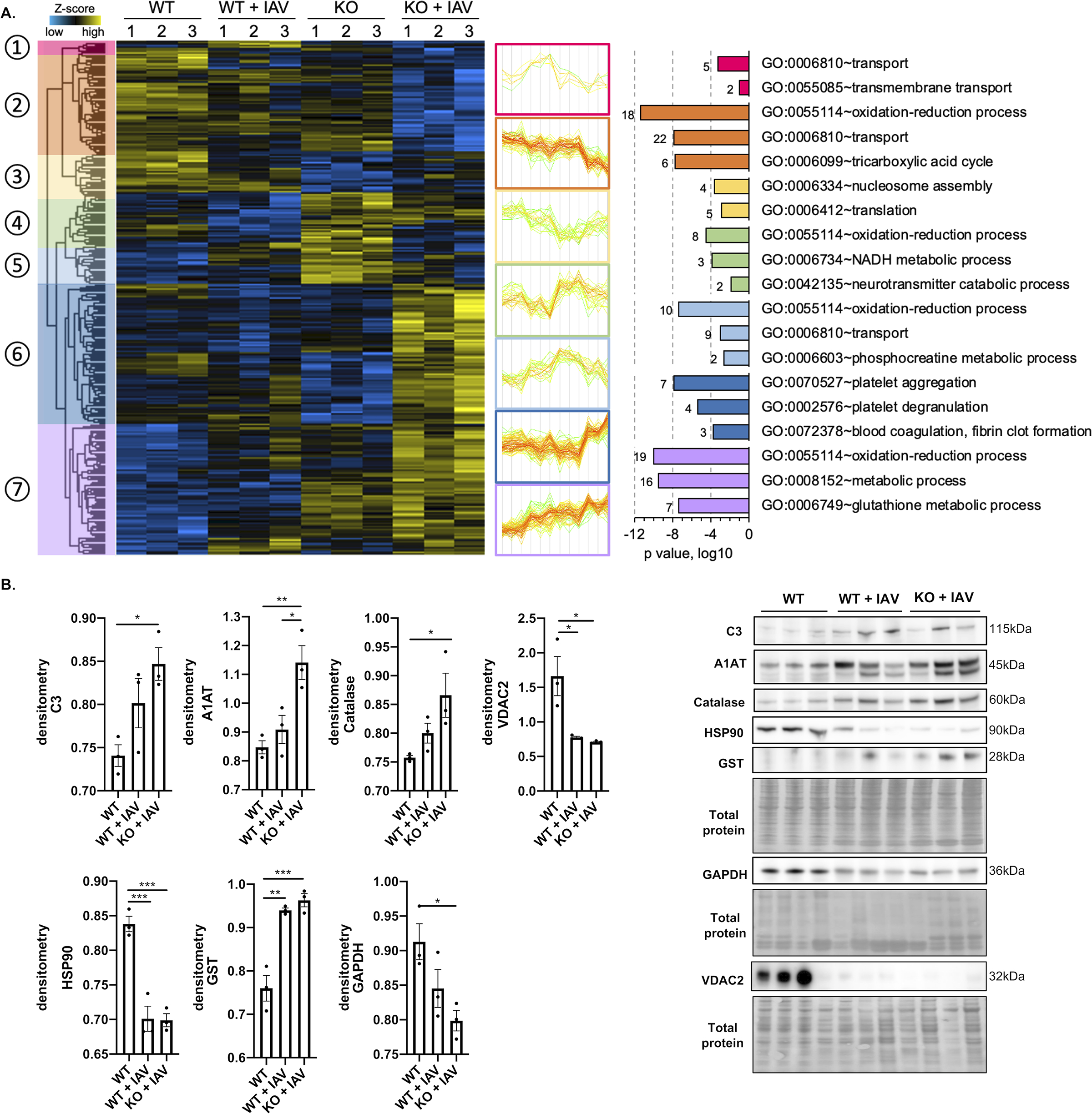
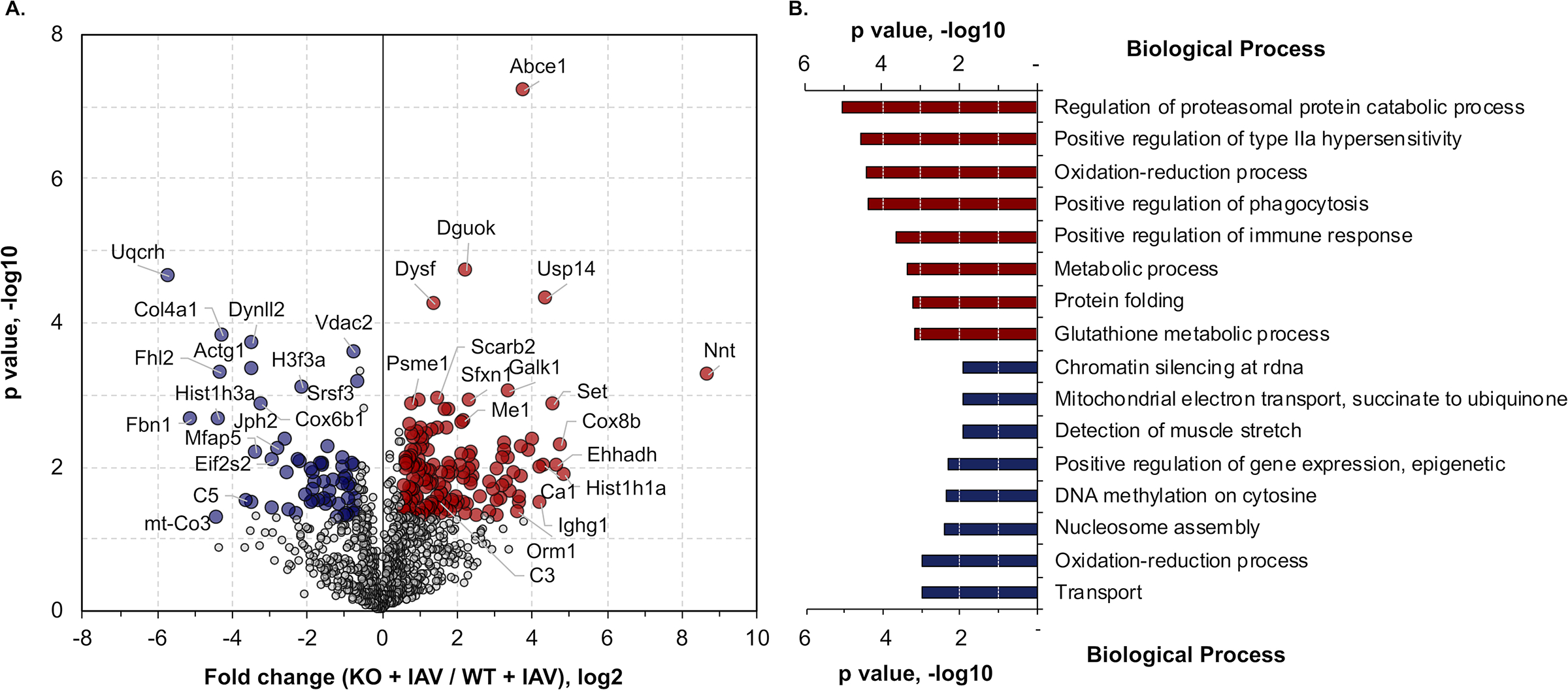
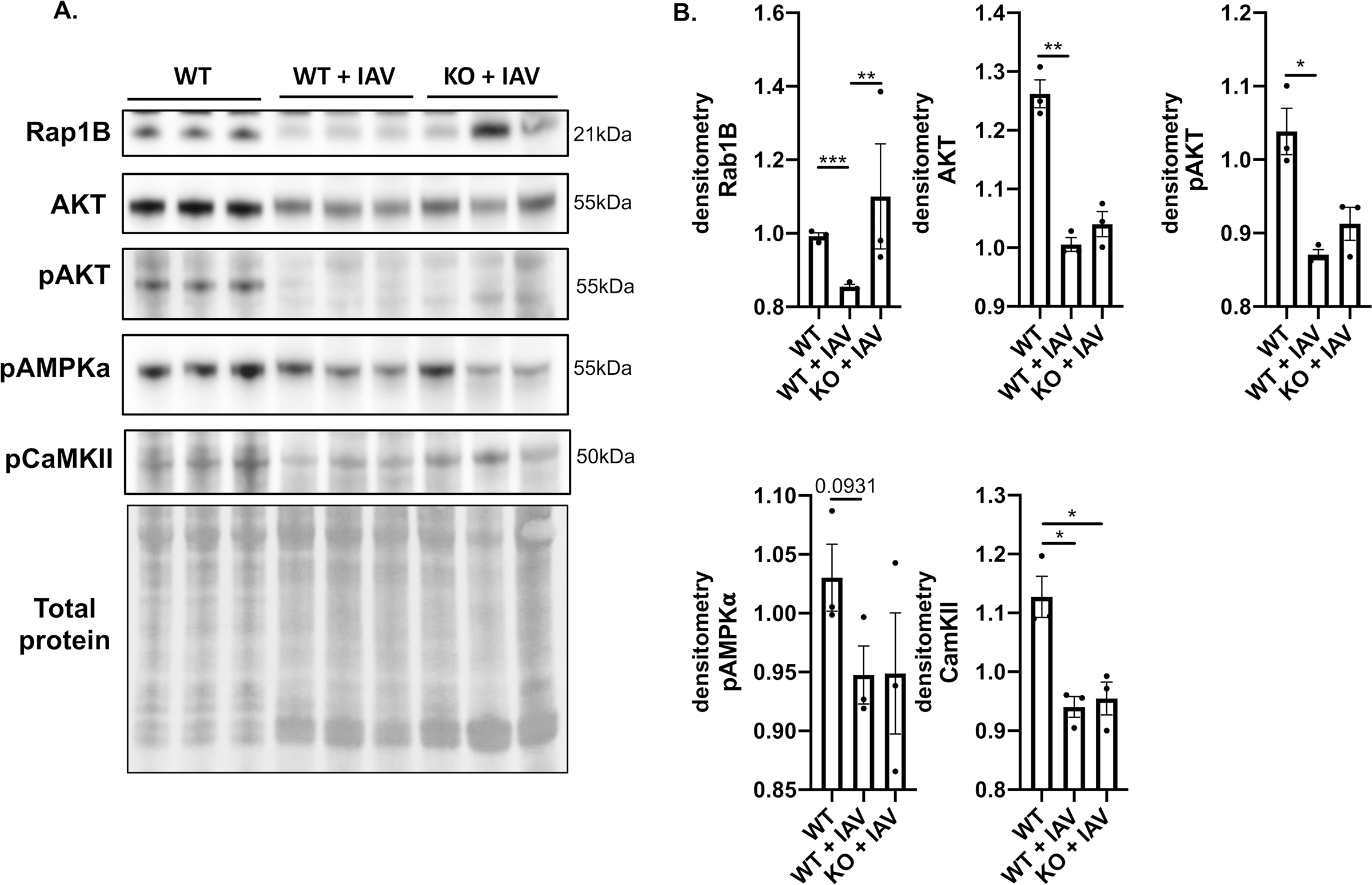
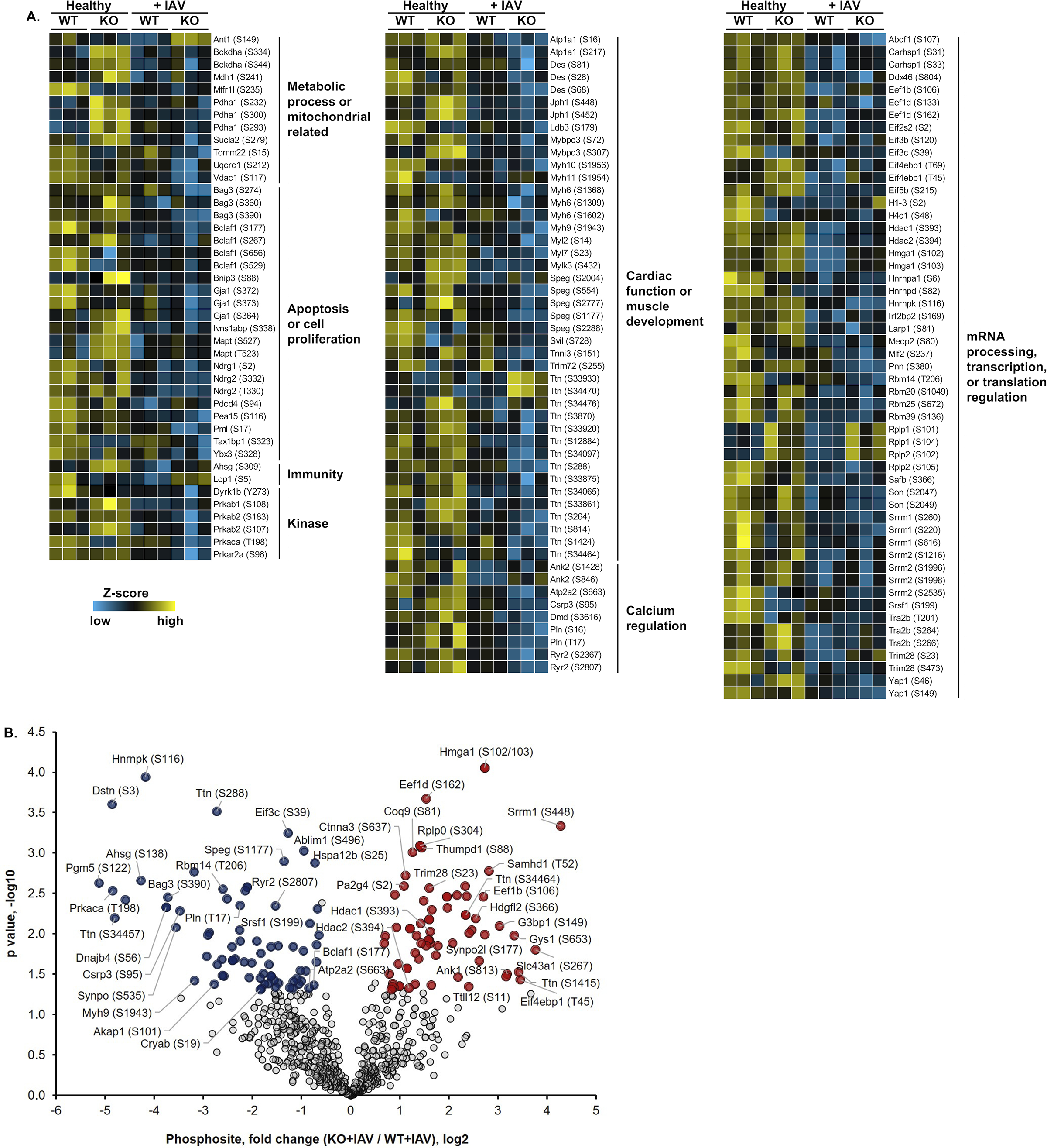
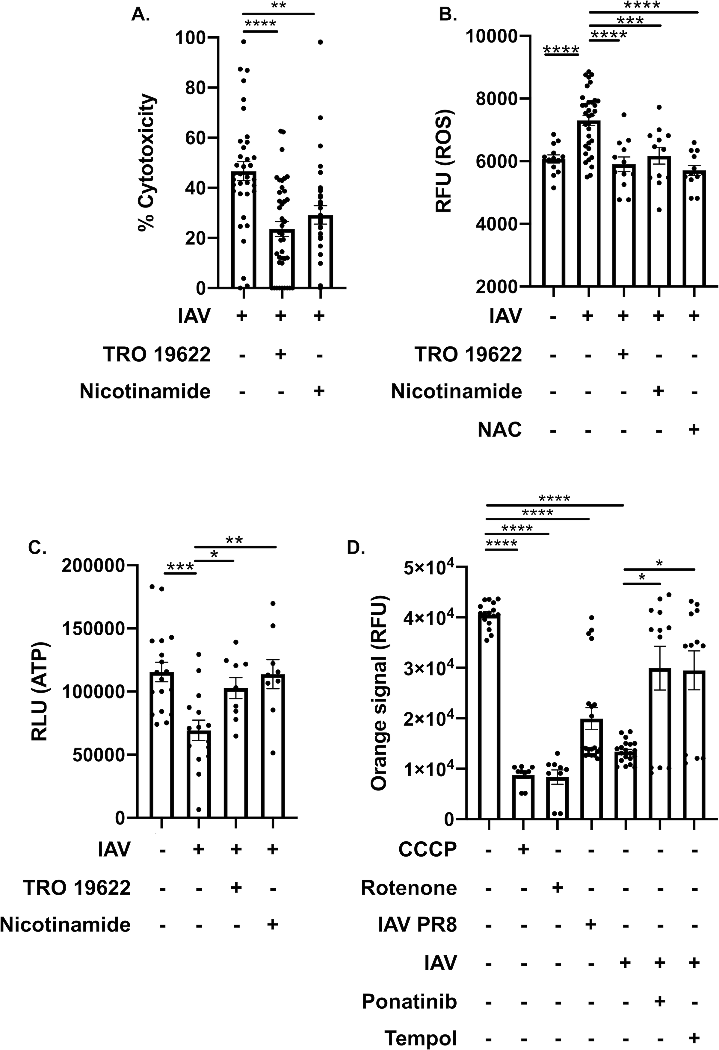
Methods and results: Hearts from wild-type and necroptosis-deficient (MLKL [mixed lineage kinase domain-like protein]-KO) mice were dissected 12 days after initial influenza A virus (IAV) infection when viral titers were undetectable in the lungs. Immunofluorescence microscopy and plaque assays showed presence of viable IAV particles in the myocardium without generation of interferon responses. Global proteome and phosphoproteome analyses using high-resolution accurate mass-based LC-MS/MS and label-free quantitation showed that the global proteome as well as the phosphoproteome profiles were significantly altered in IAV-infected mouse hearts in a strain-independent manner. Necroptosis-deficient mice had increased survival and reduced weight loss post-IAV infection, as well as increased antioxidant and mitochondrial function, indicating partial protection to IAV infection. These findings were confirmed in vitro by pretreatment of human and rat myocytes with antioxidants or necroptosis inhibitors, which blunted oxidative stress and mitochondrial damage after IAV infection.

Conclusions: This study provides the first evidence that the cardiac proteome and phosphoproteome are significantly altered post-pulmonary influenza infection. Moreover, viral particles can persist in the heart after lung clearance, altering mitochondrial function and promoting cell death without active replication and interferon responses. Finally, our findings show inhibition of necroptosis or prevention of mitochondrial damage as possible therapeutic interventions to reduce cardiac damage during influenza infections. Graphic Abstract: A graphic abstract is available for this article.

Keywords: cell death; heart; human influenza; mitochondria; necroptosis; oxidative stress; proteomics.


Figures

Figure 1:
Figure 1:. Influenza A virus infection leads to programmed necrosis, oxidative stress and metabolic breakdown in cardiomyocytes.
(A) LDH release of H9c2 rat myocytes were infected with an MOI of 0.1 of IAV strain Influenza A virus/California/7/2009 (IAV) for 48-h. (B) Immunofluorescent staining for pMLKL (green) in H9c2 rat myocytes infected with IAV. Cell nucleus was stained in blue. White bar = 50 μm. (C) Mean fluorescence intensity (MFI) of pMLKL. (D) LDH cytotoxicity assay of H9c2 infected with IAV at an MOI of 0.1 for 48-h. Cells were treated with two necroptosis inhibitors: ponatinib and pazopanib (10 μM each) for 1h before infection. (E) Propidium iodide and (F) LDH cytotoxicity assay staining of human AC16 myocytes infected with IAV at an MOI of 0.1 for 48-h. Cells were treated with MLKL inhibitor necrosulfonamide (10 μM each) for 1h before infection. (G) 8-oxoguanine (8-OXO, green) was stained in H9c2 rat myocytes infected with IAV. Cell nucleus was stained in blue. White bar = 50 μm. (H) Mean fluorescence intensity (MFI) of 8-OXO staining. (I) LDH cytotoxicity assay of H9c2 infected with IAV at an MOI of 0.1 for 48-h. Cells were treated with Catalase (10 μM), Tempol (10 μM), Rotenone + Thallium trifluoroacetate (Rot tha, 10 nM/mL/10 nM/mL), Allopurinol (Allu, 10 nM/mL), Apocynin (Apo, 1 μM/mL), Mefenamic acid (Mefe, 20 nM/mL). (J) ATP levels and (K) ROS/superoxide levels of IAV-infected H9c2 myocytes treated with ponatinib (10 μM), compared to mock treated and uninfected H9c2. RLU: relative light unit of luminescence. Mann-Whitney U tests were applied for two-group comparisons. Kruskal-Wallis test with Dunn’s multiple-comparison post-test. Asterisks denote the level of significance observed: * = p ≤ 0.05; ** = p ≤ 0.01; *** = p ≤ 0.001, **** = p ≤ 0.0001.
Figure 2:
Figure 2:. Influenza virus persists in mice hearts during convalescence without exacerbated inflammatory response.
(A) Timeline for mice infection and heart collection. Male and female wild-type and MLKL KO 6-to-8-week-old C57Bl/6 mice were intranasally infected with A/California/7/2009 (IAV) at day 0. Mice were euthanized and heart tissue was collected at day 12 (n = 10–12, 5–6 mice of each sex). (B) survival and (C) percent weight changes up to 10 days post IAV infection. (D) Viral titers were measured for WT C57Bl/6 and MLKL KO mice 12 days post initial IAV infection (n = 6, 3 of each sex) by the plaque assay and plotted in Log PFU/mL. (E) Levels of IFNƔ, TNF⍺, IFN⍺, IL-10, IFN-ƛ and IL-1-β measured by ELISA (pg/mL, n = 3–6, representative data for each sex of mice infected with IAV or challenged with vehicle (PBS) control). Log-rank (Mantel-Cox) test for survival. Kruskal-Wallis test with Dunn’s multiple-comparison post-test. Asterisks denote the level of significance observed: * = p ≤ 0.05; ** = p ≤ 0.01; *** = p ≤ 0.001.
Figure 3:
Figure 3:. Influenza infection induces cardiac proteome remodeling.
Male and female 6-to-8-week-old wild-type and MLKL KO C57Bl/6 mice were intranasally infected with A/California/7/2009 (IAV) or mock challenged with vehicle (PBS) and hearts excised at day 12 post-infection. (A) Proteomic changes of mice hearts after IAV infection or mock challenge (n = 3 per condition). Hierarchical clustering of LFQ intensities of 295 significantly changed proteins (ANOVA, FDR < 0.01) revealed seven distinct clusters. Their abundance profiles among the groups were plotted in the middle panel. Enriched GO biological process terms are indicated for each marked cluster on the right panel. (B) Immunoblots for complement C3 (C3), AIAT, Catalase, HSP90, GST, GAPDH and VDAC and histograms of protein level quantification (n = 3 per group). Proteomic data of mice is representative from 2 separate experiments done with 3 mice of each sex; no sex based differences were observed. Kruskal-Wallis test with Dunn’s multiple-comparison post-test. Asterisks denote the level of significance observed: * = p ≤ 0.05; ** = p ≤ 0.01; *** = p ≤ 0.001.
Figure 4:
Figure 4:. MLKL deficiency leads to proteome reshape in mice hearts during convalescence.
(A) Quantitative comparison of IAV infected WT and MLKL KO mice (n = 3 per group). Fold change of the proteins (x-axis) and their significance (p value, y-axis) were plotted. Up- and down-regulated proteins (1.5-fold cutoff) were highlighted in red and blue, respectively. (B) GO enrichment analysis of the significantly changed proteins as shown in panel A. Top 8 enriched biological terms of each group were displayed. Proteomic data of control and infected mice is representative from 2 separate experiments done with 3 mice of each sex; no sex based differences were observed.
Figure 5:
Figure 5:. Influenza infection leads to downregulation of functional proteins in the heart.
Male and female 6-to-8-week-old wild-type and MLKL KO mice were intranasally infected with A/California/7/2009 (IAV) or mock challenged with vehicle (PBS) and hearts excised at day 12 post-infection. (n = 3 per group, representative of 2 experiments for a total n =6, with 3 mice of each sex). Heart tissue was collected and immunoblots were performed against Rap1B, AKT, pAKT, pAMPKa, and pCaMKII, presented with quantification of relative protein level. Kruskal-Wallis test with Dunn’s multiple-comparison post-test. Asterisks denote the level of significance observed: * = p ≤ 0.05; ** = p ≤ 0.01.
Figure 6:
Figure 6:. Influenza virus infection leads to changes in phosphorylation of cardiac proteins.
(A) Quantitative phosphoproteomic profiling of mouse heart. Plotted here are selected proteins and their identified phosphosites (indicated in the parentheses) grouped by protein functions or related pathways. (B) Quantitative comparison of IAV infected or mock infected (PBS challenge) WT and MLKL KO mice (n = 3 per group). Fold change of the phosphosites (x-axis) and their significance (p value, y-axis) were plotted. Up- and down-regulated proteins (1.5-fold cutoff) are highlighted in red and blue, respectively. Phosphoproteomic data is representative from 2 separate experiments done with 3 mice of each sex, no sex based differences were observed.
Figure 7:
Figure 7:. Inhibition of the MPTP and replenishment of NAD+ leads to cardiomyocyte protection after IAV infection.
(A) Cytotoxicity of H9c2 myocytes treated with TRO 19622 (10 μM) or nicotinamide (1 mM) infected with A/California/7/2009 (IAV). (B) ROS/superoxide levels and (C) ATP levels of IAV-infected H9c2 myocytes treated with TRO 19622, nicotinamide with same concentration in (A) or n-acetylcysteine (10 μM), compared to uninfected or mock treated H9c2. RLU: relative light unit of luminescence. (D) Mitochondrial membrane permeabilization assay as measured via changes in RFU: relative fluorescent units of human AC16 cardiomyocytes infected with A/California/7/2009 (IAV) or PR8 and treated with rotenone, tempol or ponatinib. Carbonyl cyanide m-chlorophenyl hydrazone (CCCP) positive control for mitochondrial membrane permeabilization. Kruskal-Wallis test with Dunn’s multiple-comparison post-test. Asterisks denote the level of significance observed: * = p ≤ 0.05; ** = p ≤ 0.01; *** = p ≤ 0.001, **** = p ≤ 0.0001.

Similar articles

Cited by

References

    1. Mortality GBD, Causes of Death C. Global, regional, and national life expectancy, all-cause mortality, and cause-specific mortality for 249 causes of death, 1980–2015: A systematic analysis for the global burden of disease study 2015. Lancet. 2016;388:1459–1544 - PMC - PubMed
    1. Heron M Deaths: Leading causes for 2017. National vital statistics reports. 2019;68 - PubMed
    1. Paules C, Subbarao K. Influenza. Lancet. 2017;390:697–708 - PubMed
    1. Noda T [orthomyxoviruses]. Uirusu. 2012;62:219–228 - PubMed
    1. Thompson WW, Shay DK, Weintraub E, Brammer L, Cox N, Anderson LJ, Fukuda K. Mortality associated with influenza and respiratory syncytial virus in the united states. JAMA. 2003;289:179–186 - PubMed

Publication types

MeSH terms