

Granadaene Photobleaching Reduces the Virulence and Increases Antimicrobial Susceptibility of Streptococcus agalactiae
- PMID: 33502005
- PMCID: PMC8277675
- DOI: 10.1111/php.13389
Granadaene Photobleaching Reduces the Virulence and Increases Antimicrobial Susceptibility of Streptococcus agalactiae
Abstract
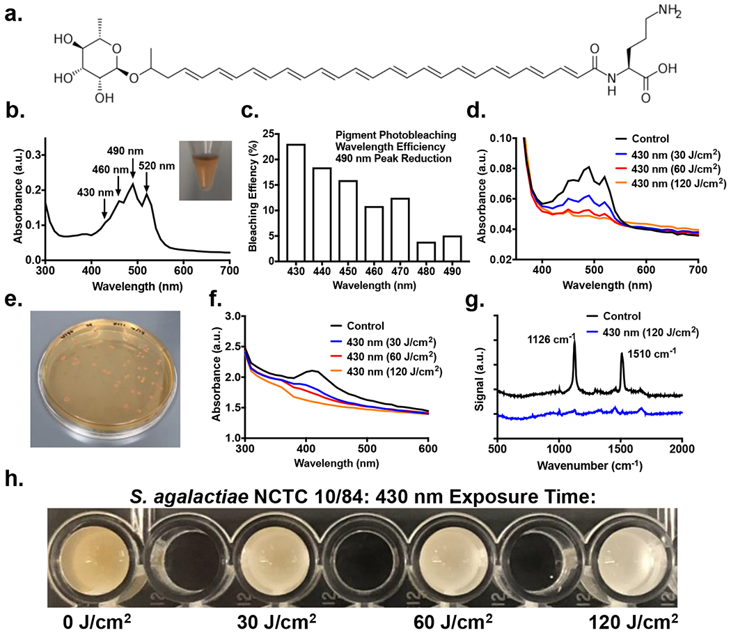
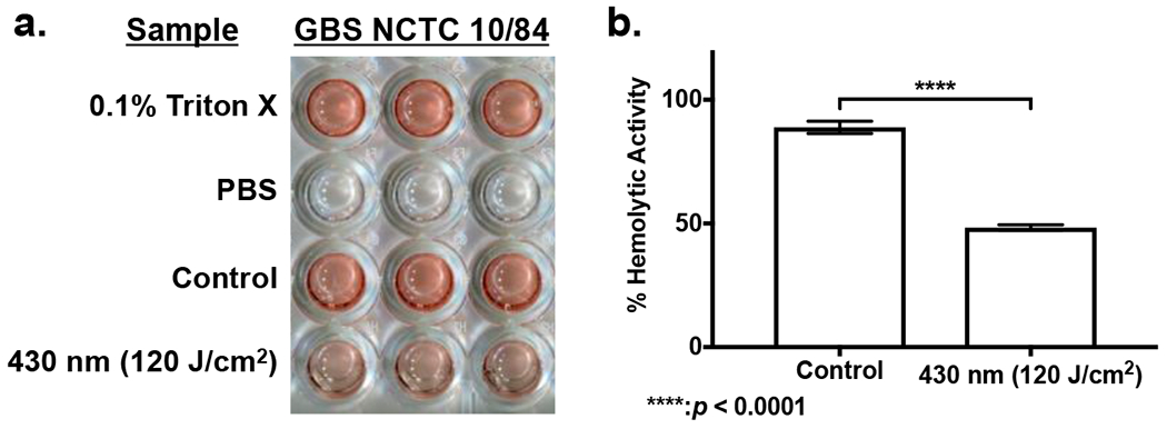
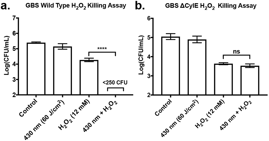
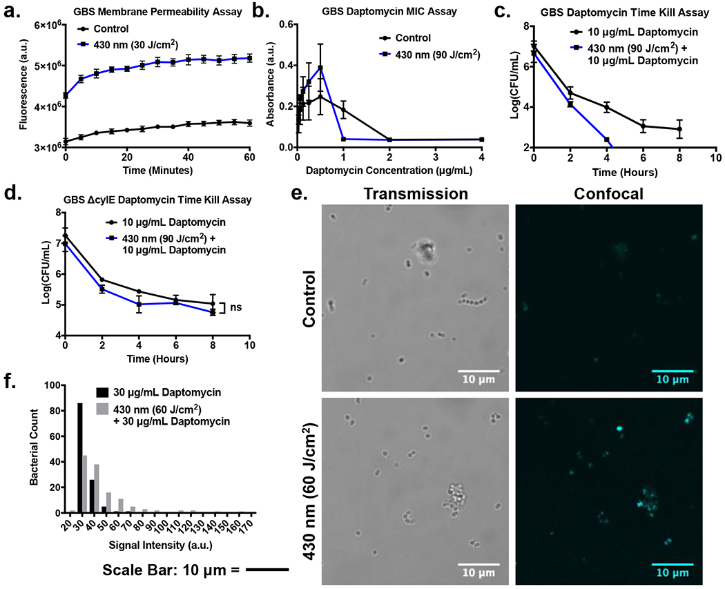
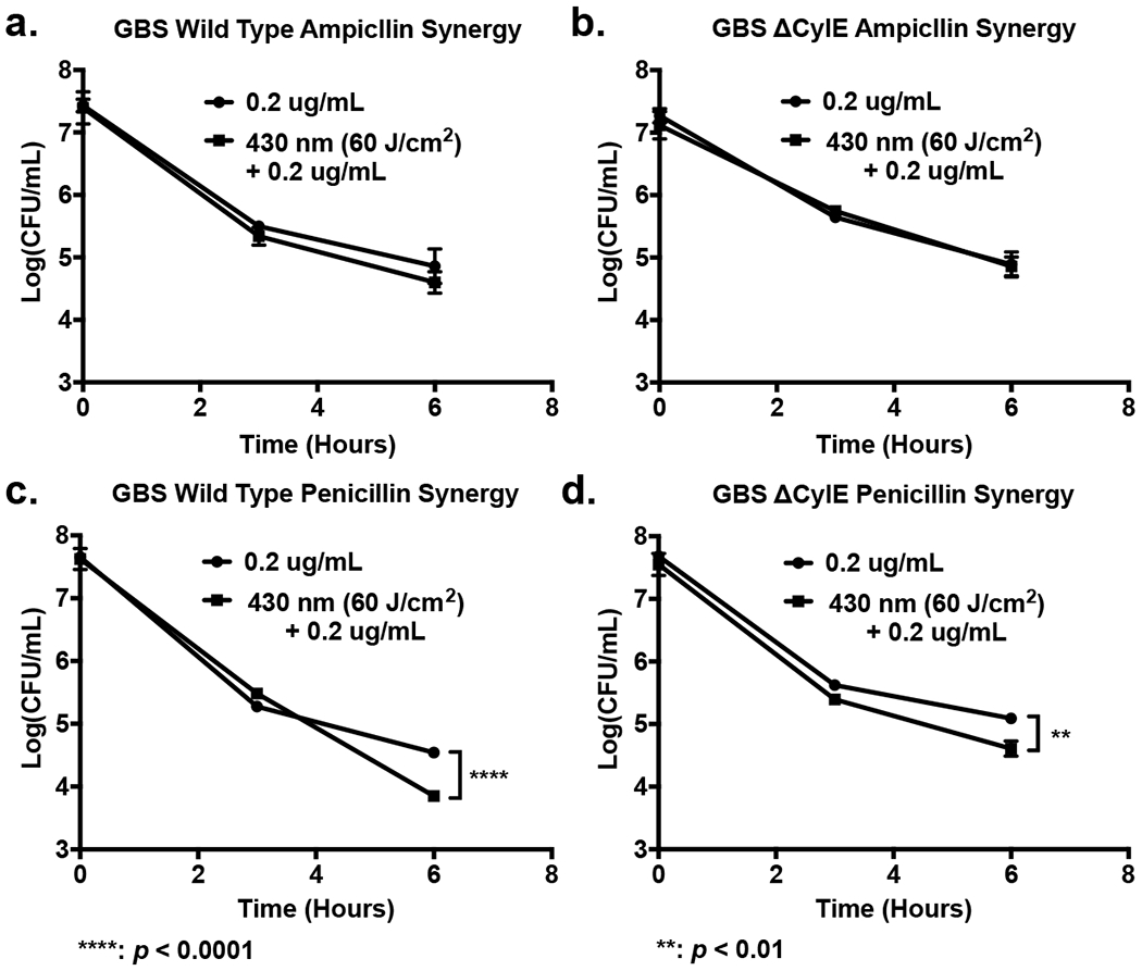
Streptococcus agalactiae, also known as Group B Streptococcus (GBS), is increasingly recognized as a major cause of soft tissue and invasive diseases in the elderly and diabetic populations. Antibiotics like penicillin are used with great frequency to treat these infections, although antimicrobial resistance is increasing among GBS strains and underlines a need for alternative methods not reliant on traditional antibiotics. GBS granadaene pigment is related to the hemolysin/cytolysin of GBS, which is critical for the pathogenesis of GBS diseases. Here, we show that photobleaching granadaene dampens the hemolytic activity of GBS. Furthermore, photobleaching of this antioxidant was found to increase GBS susceptibility to killing by reactive oxygen species like hydrogen peroxide. Treatment with light was also shown to affect GBS membrane permeability and contribute to increased susceptibility to the cell membrane-targeting antibiotic daptomycin. Overall, our study demonstrates dual effects of photobleaching on the virulence and antimicrobial susceptibility of GBS and suggests a novel approach for the treatment of GBS infection.
© 2021 American Society for Photobiology.
Figures
Similar articles
-
Hemolytic Membrane Vesicles of Group B Streptococcus Promote Infection.J Infect Dis. 2021 Apr 23;223(8):1488-1496. doi: 10.1093/infdis/jiaa548. J Infect Dis. 2021. PMID: 32861213 Free PMC article.
-
Molecular Characteristics of IS1216 Carrying Multidrug Resistance Gene Cluster in Serotype III/Sequence Type 19 Group B Streptococcus.mSphere. 2021 Aug 25;6(4):e0054321. doi: 10.1128/mSphere.00543-21. Epub 2021 Jul 28. mSphere. 2021. PMID: 34319128 Free PMC article.
-
Group B streptococcal haemolysin and pigment, a tale of twins.FEMS Microbiol Rev. 2014 Sep;38(5):932-46. doi: 10.1111/1574-6976.12071. Epub 2014 Apr 4. FEMS Microbiol Rev. 2014. PMID: 24617549 Free PMC article. Review.
-
Antibiotic resistance, biofilm formation, and virulence genes of Streptococcus agalactiae serotypes of Indian origin.BMC Microbiol. 2023 Jul 5;23(1):176. doi: 10.1186/s12866-023-02877-y. BMC Microbiol. 2023. PMID: 37407919 Free PMC article.
-
A review of antibiotic resistance in Group B Streptococcus: the story so far.Crit Rev Microbiol. 2020 May;46(3):253-269. doi: 10.1080/1040841X.2020.1758626. Epub 2020 May 2. Crit Rev Microbiol. 2020. PMID: 32363979 Review.
Cited by
-
An opportunistic pathogen under stress: how Group B Streptococcus responds to cytotoxic reactive species and conditions of metal ion imbalance to survive.FEMS Microbiol Rev. 2024 May 8;48(3):fuae009. doi: 10.1093/femsre/fuae009. FEMS Microbiol Rev. 2024. PMID: 38678005 Free PMC article. Review.
-
Characterization of AI-2/LuxS quorum sensing system in biofilm formation, pathogenesis of Streptococcus equi subsp. zooepidemicus.Front Cell Infect Microbiol. 2024 Feb 6;14:1339131. doi: 10.3389/fcimb.2024.1339131. eCollection 2024. Front Cell Infect Microbiol. 2024. PMID: 38379770 Free PMC article.
-
Photoinactivation of Catalase Sensitizes Candida albicans and Candida auris to ROS-Producing Agents and Immune Cells.Adv Sci (Weinh). 2022 Apr;9(10):e2104384. doi: 10.1002/advs.202104384. Epub 2022 Feb 4. Adv Sci (Weinh). 2022. PMID: 35119220 Free PMC article.
-
Chromophore-Targeting Precision Antimicrobial Phototherapy.Cells. 2023 Nov 20;12(22):2664. doi: 10.3390/cells12222664. Cells. 2023. PMID: 37998399 Free PMC article. Review.
-
Group B Streptococcus: Virulence Factors and Pathogenic Mechanism.Microorganisms. 2022 Dec 15;10(12):2483. doi: 10.3390/microorganisms10122483. Microorganisms. 2022. PMID: 36557736 Free PMC article. Review.
References
-
- Török E and Day N (2005) Staphylococcal and streptococcal infections. Medicine 33, 97–100. 10.1383/medc.33.5.97.64964. - DOI
Publication types
MeSH terms
Substances
Grants and funding
LinkOut - more resources
Full Text Sources
Other Literature Sources
Medical
Molecular Biology Databases
