. 2021 Feb;7(2):000514.
doi: 10.1099/mgen.0.000514.

Colonization with Staphylococcus aureus and Klebsiella pneumoniae causes infections in a Vietnamese intensive care unit

Affiliations

Colonization with Staphylococcus aureus and Klebsiella pneumoniae causes infections in a Vietnamese intensive care unit

Duong Bich Thuy et al. Microb Genom. 2021 Feb.

Abstract

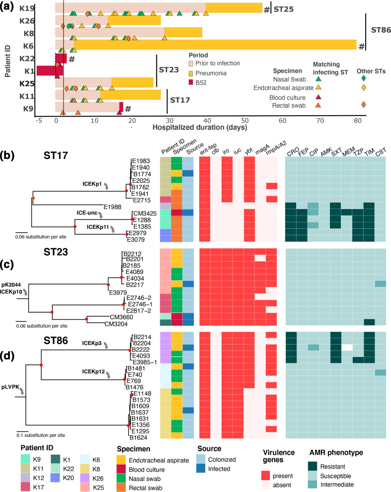
Pre-existing colonization with Staphylococcus aureus or Klebsiella pneumoniae has been found to increase the risk of infection in intensive care patients. We previously conducted a longitudinal study to characterize colonization of these two organisms in patients admitted to intensive care in a hospital in southern Vietnam. Here, using genomic and phylogenetic analyses, we aimed to assess the contribution these colonizing organisms made to infections. We found that in the majority of patients infected with S. aureus or K. pneumoniae, the sequence type of the disease-causing (infecting) isolate was identical to that of corresponding colonizing organisms in the respective patient. Further in-depth analysis revealed that in patients infected by S. aureus ST188 and by K. pneumoniae ST17, ST23, ST25 and ST86, the infecting isolate was closely related to and exhibited limited genetic variation relative to pre-infection colonizing isolates. Multidrug-resistant S. aureus ST188 was identified as the predominant agent of colonization and infection. Colonization and infection by K. pneumoniae were characterized by organisms with limited antimicrobial resistance profiles but extensive repertoires of virulence genes. Our findings augment the understanding of the link between bacterial colonization and infection in a low-resource setting, and could facilitate the development of novel evidence-based approaches to prevent and treat infections in high-risk patients in intensive care.

Keywords: Staphylococcus aureus; colonization; hospital-acquired infections; hypervirulent Klebsiella pneumoniae; intra-patient diversity.


Conflict of interest statement

The authors declare that there are no conflict of interests.

Figures

Fig. 1.
Fig. 1.
Schematic graph showing the colonization and infection of (a) 19 patients infected by Staphylococcus aureus and (b) 28 patients infected by Klebsiella pneumoniae . For colonization upon admission or acquired during ICU stay, each square box denotes a positive (coloured) or negative (grey) culture result from the patient’s respective body sites (as seen in the key). Missing boxes indicate a loss of surveillance culture during ICU stay. For infection, the colour of each box indicates the nature of disease (see key), with black-lined boxes denoting missing infecting isolates. Black and red triangles denote the methicillin resistance and extreme-drug resistance status in S. aureus and K. pneumoniae infections, respectively. A filled line connecting the boxes in each patient indicates that the infecting and colonizing isolates are of the same ST, with pink and blue lines showing that infections are caused by ST188 S. aureus and S. argenteus respectively. Patient IDs with blue shading indicate that the patient died due to the infection during the ICU stay.
Fig. 2.
Fig. 2.
Genomic investigation of S. aureus ST188 causing infections in this study. (a) Schematic representation of patients’ ICU stay, with the bar colour corresponding to the infection status during or prior to the ICU stay (see key). Each triangle represents a positive culture of ST188 S. aureus from the patient at defined time point since ICU admission, with colour matching the type of clinical specimen (see key). Each filled diamond represents a positive non-ST188 S. aureus culture from the respective specimen (as coloured in the key). A ‘#’ symbol denotes that the respective patient died during the ICU stay. A solid vertical black line denotes the time point of 48 h after hospital admission. (b) Maximum-likelihood phylogeny of 39 ST188 S. aureus isolated in this study. The tree is mid-point rooted, and red filled circles indicate bootstrap values >80 at the internal nodes. The columns show the associated data for each taxon, including Patient ID, specimen and source of isolation (colonizing or infecting). Grey arrows denote the acquisition of accessory genetic elements into the phylogeny. (c) Dfferences (in SNPs) of ST188 S. aureus isolated within each patient (excluding S10 and S19), as assessed by reference-based recombination-free mapping. Each dot represents an SNP difference between two isolates, and the boxplot shows the distribution of such differences in each patient.
Fig. 3.
Fig. 3.
Genomic investigation of major K. pneumoniae STs causing infections in this study. (a) Schematic representation of patients’ ICU stay, with the bar colour corresponding to the infection status during or prior to ICU stay (see key). The vertical black line includes patients infected by the respective K. pneumoniae ST (ST17, ST23, ST86, ST25). Each triangle represents a positive culture of K. pneumoniae whose ST matches that of the infecting isolate, with colour denoting the type of clinical specimen (see key). Each filled diamond represents a positive K. pneumoniae culture of STs not matching that of the infecting isolate, from the specimen as coloured in the key. A ‘#’ symbol denotes that the respective patient died during the ICU stay. A solid vertical black line denotes the time point of 48 h after hospital admission. The remaining panels display the maximum-likelihood phylogeny of (b) ST17, (c) ST23 and (d) ST86 K. pneumoniae isolated from this study. Each tree is mid-point rooted, and red filled circles indicate bootstrap values >80 at the internal nodes. The columns show the associated information for each taxon, including Patient ID, specimen and source of isolation (colonizing or infecting); the presence of virulence genes, including ent-fep (enterobactin), clb (colibactin), iro (salmochelin), iuc (aerobactin), ybt (yersiniabactin), magA (mucoviscosity associated gene A), rmpA/A2 (regulator of mucoid phenotype A/A2); susceptibility to antimicrobials, including CRO (ceftriaxone), FEP (cefepime), CIP (ciprofloxacin), AMK (amikacin), SXT (co-trimoxazole), MEM (meropenem), TZP (piperacillin/tazobactam), TIM (ticarcillin/clavulanate), CST (colistin). Grey arrows denote the acquisition of accessory genetic elements into the phylogeny. ICE-unc: uncharacterized ICE.

Similar articles

Cited by

References

    1. Donowitz LG, Wenzel RP, Hoyt JW. High risk of hospital-acquired infection in the ICU patient. Crit Care Med. 1982;10:355–357. doi: 10.1097/00003246-198206000-00001. p. - DOI - PubMed
    1. Weber DJ, Raasch R, Rutala WA. Nosocomial infections in the ICU: the growing importance of antibiotic-resistant pathogens. Chest. 1999;115:34S–41. doi: 10.1378/chest.115.suppl_1.34s. - DOI - PubMed
    1. WHO Report on the burden of endemic health care-associated infection worldwide clean care is safer care. World Heal Organ. 2011:1–40.
    1. Baker S, Thomson N, Weill FX, Holt KE. Genomic insights into the emergence and spread of antimicrobial-resistant bacterial pathogens. Science. 2018;360:733–738. doi: 10.1126/science.aar3777. - DOI - PMC - PubMed
    1. Honda H, Krauss MJ, Coopersmith CM, Kollef MH, Richmond AM, et al. Staphylococcus aureus nasal colonization and subsequent infection in intensive care unit patients: does methicillin resistance matter? Infect Control Hosp Epidemiol. 2010;31:584–591. doi: 10.1086/652530. - DOI - PMC - PubMed

Publication types

MeSH terms

LinkOut - more resources