

ATP activation of peritubular cells drives testicular sperm transport
- PMID: 33502316
- PMCID: PMC7840184
- DOI: 10.7554/eLife.62885
ATP activation of peritubular cells drives testicular sperm transport
Abstract
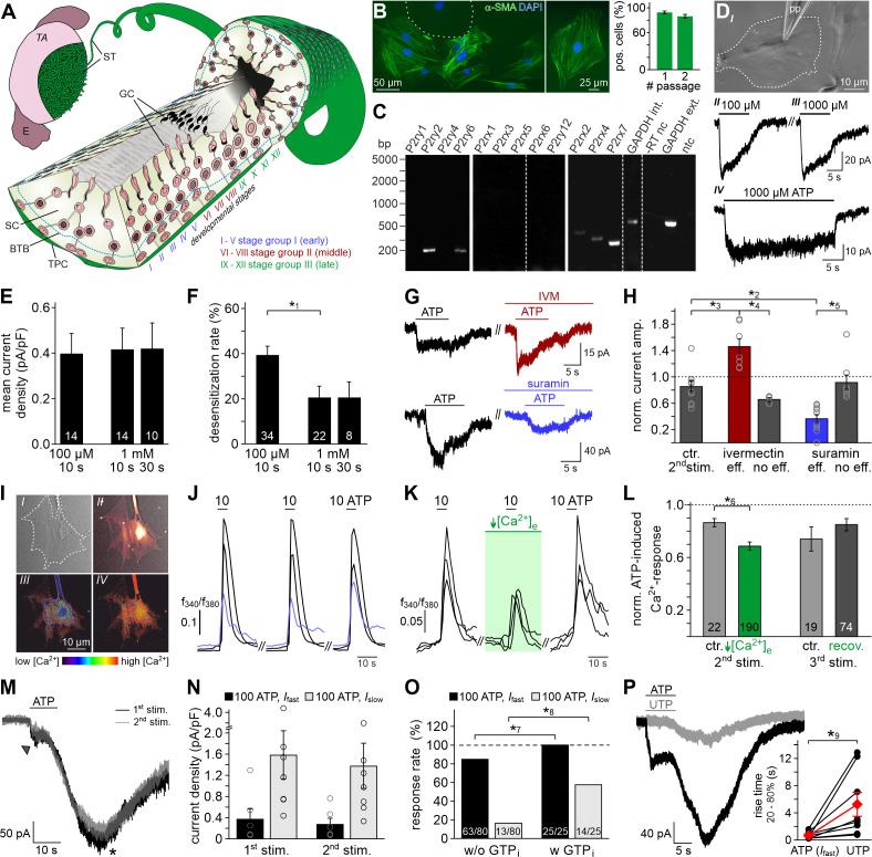
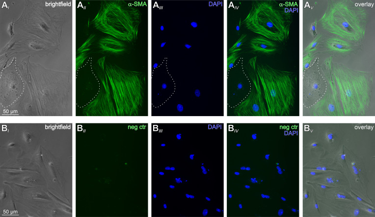
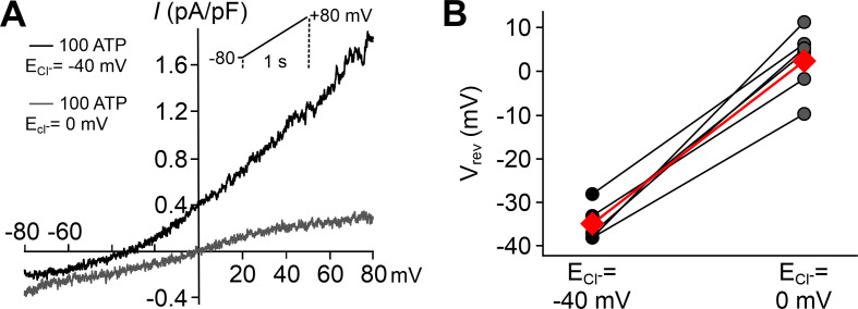
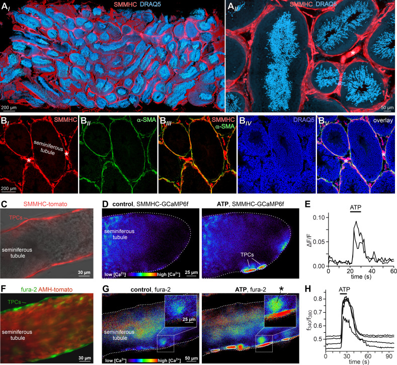
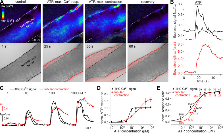
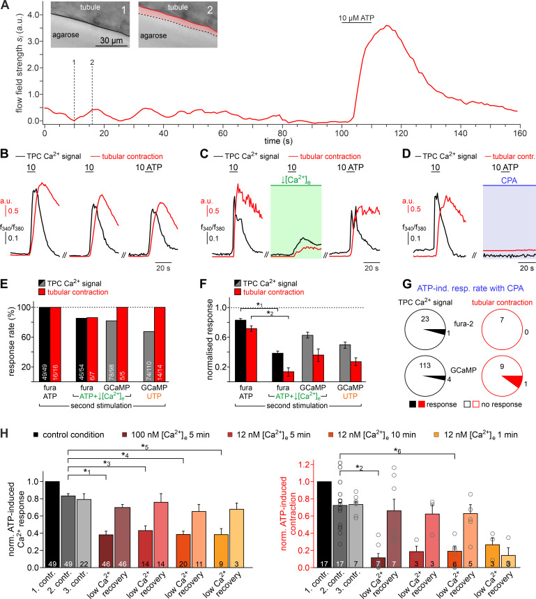
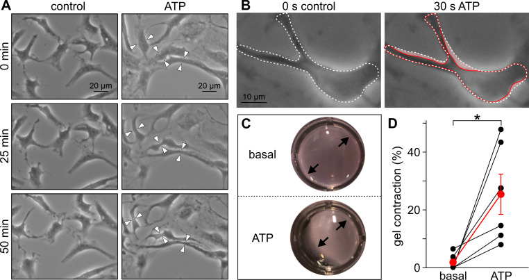
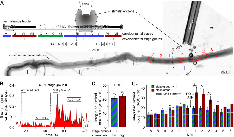
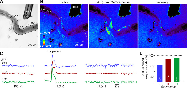
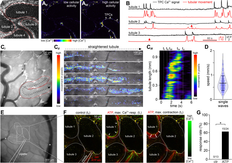
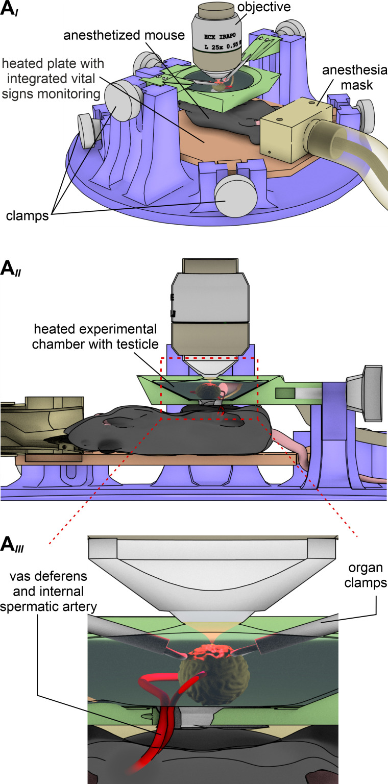
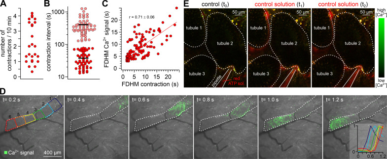
Spermatogenesis, the complex process of male germ cell proliferation, differentiation, and maturation, is the basis of male fertility. In the seminiferous tubules of the testes, spermatozoa are constantly generated from spermatogonial stem cells through a stereotyped sequence of mitotic and meiotic divisions. The basic physiological principles, however, that control both maturation and luminal transport of the still immotile spermatozoa within the seminiferous tubules remain poorly, if at all, defined. Here, we show that coordinated contractions of smooth muscle-like testicular peritubular cells provide the propulsive force for luminal sperm transport toward the rete testis. Using a mouse model for in vivo imaging, we describe and quantify spontaneous tubular contractions and show a causal relationship between peritubular Ca2+ waves and peristaltic transport. Moreover, we identify P2 receptor-dependent purinergic signaling pathways as physiological triggers of tubular contractions both in vitro and in vivo. When challenged with extracellular ATP, transport of luminal content inside the seminiferous tubules displays stage-dependent directionality. We thus suggest that paracrine purinergic signaling coordinates peristaltic recurrent contractions of the mouse seminiferous tubules to propel immotile spermatozoa to the rete testis.
Keywords: P2 receptors; cell biology; germ cell development; human; male reproduction; mouse; purinergic signaling; spermatogenesis.
Plain language summary
As sperm develop in the testis, the immature cells must make their way through a maze of small tubes known as seminiferous tubules. However, at this stage, the cells do not yet move the long tails that normally allow them to ‘swim’; it is therefore unclear how they are able to move through the tubules. Now, Fleck, Kenzler et al. have showed that, in mice, muscle-like cells within the walls of seminiferous tubules can create waves of contractions that push sperm along. Further experiments were then conducted on cells grown in the laboratory. This revealed that a signaling molecule called ATP orchestrates the moving process by activating a cascade of molecular events that result in contractions. Fleck, Kenzler et al. then harnessed an advanced microscopy technique to demonstrate that this mechanism occurs in living mice. Together, these results provide a better understanding of how sperm mature, which could potentially be relevant for both male infertility and birth control.
© 2021, Fleck et al.
Conflict of interest statement
DF, LK, NM, MS, NU, RM, FB, AM, AM, DM, JS, MS No competing interests declared
Figures
Similar articles
-
Three-dimensional analysis and in vivo imaging for sperm release and transport in the murine seminiferous tubule.Reproduction. 2022 May 23;164(1):9-18. doi: 10.1530/REP-21-0400. Reproduction. 2022. PMID: 35521906
-
Contractility of seminiferous tubules as related to sperm transport in the male.Arch Androl. 1981 Jun;6(4):283-94. doi: 10.3109/01485018108987539. Arch Androl. 1981. PMID: 6113819 Review.
-
Irradiation affects germ and somatic cells in prepubertal monkey testis xenografts.Mol Hum Reprod. 2017 Mar 1;23(3):141-154. doi: 10.1093/molehr/gax003. Mol Hum Reprod. 2017. PMID: 28130393
-
Transplantation of testis germinal cells into mouse seminiferous tubules.Int J Dev Biol. 1997 Feb;41(1):111-22. Int J Dev Biol. 1997. PMID: 9074943
-
Human testicular peritubular cells: more than meets the eye.Reproduction. 2013 Apr 29;145(5):R107-16. doi: 10.1530/REP-12-0497. Print 2013 May. Reproduction. 2013. PMID: 23431272 Review.
Cited by
-
Age-Related Alterations in the Testicular Proteome of a Non-Human Primate.Cells. 2021 May 24;10(6):1306. doi: 10.3390/cells10061306. Cells. 2021. PMID: 34074003 Free PMC article.
-
Dynamic volumetric imaging and cilia beat mapping in the mouse male reproductive tract with optical coherence tomography.Biomed Opt Express. 2022 Jun 1;13(6):3672-3684. doi: 10.1364/BOE.459937. eCollection 2022 Jun 1. Biomed Opt Express. 2022. PMID: 35781970 Free PMC article.
-
Profound Effects of Dexamethasone on the Immunological State, Synthesis and Secretion Capacity of Human Testicular Peritubular Cells.Cells. 2022 Oct 9;11(19):3164. doi: 10.3390/cells11193164. Cells. 2022. PMID: 36231125 Free PMC article.
-
Perfusion in Organ-on-Chip Models and Its Applicability to the Replication of Spermatogenesis In Vitro.Int J Mol Sci. 2022 May 12;23(10):5402. doi: 10.3390/ijms23105402. Int J Mol Sci. 2022. PMID: 35628214 Free PMC article. Review.
-
Insights on Platelet-Derived Growth Factor Receptor α-Positive Interstitial Cells in the Male Reproductive Tract.Int J Mol Sci. 2024 Apr 8;25(7):4128. doi: 10.3390/ijms25074128. Int J Mol Sci. 2024. PMID: 38612936 Free PMC article. Review.
References
-
- Albrecht M, Rämsch R, Köhn FM, Schwarzer JU, Mayerhofer A. Isolation and cultivation of human testicular peritubular cells: a new model for the investigation of fibrotic processes in the human testis and male infertility. The Journal of Clinical Endocrinology & Metabolism. 2006;91:1956–1960. doi: 10.1210/jc.2005-2169. - DOI - PubMed
-
- Alexander SPH, Christopoulos A, Davenport AP, Kelly E, Mathie A, Peters JA, Veale EL, Armstrong JF, Faccenda E, Harding SD, Pawson AJ, Sharman JL, Southan C, Davies JA, CGTP Collaborators The concise guide to pharmacology 2019/20: g protein-coupled receptors. British Journal of Pharmacology. 2019a;176:S21–S141. doi: 10.1111/bph.14748. - DOI - PMC - PubMed
-
- Alexander SPH, Mathie A, Peters JA, Veale EL, Striessnig J, Kelly E, Armstrong JF, Faccenda E, Harding SD, Pawson AJ, Sharman JL, Southan C, Davies JA, CGTP Collaborators The concise guide to pharmacology 2019/20: ion channels. British Journal of Pharmacology. 2019b;176:S142–S228. doi: 10.1111/bph.14749. - DOI - PMC - PubMed
Publication types
MeSH terms
Substances
LinkOut - more resources
Full Text Sources
Other Literature Sources
Molecular Biology Databases
Research Materials
Miscellaneous
