Regional Differences and Physiologic Behaviors in Peripapillary Scleral Fibroblasts
- PMID: 33502460
- PMCID: PMC7846956
- DOI: 10.1167/iovs.62.1.27
Regional Differences and Physiologic Behaviors in Peripapillary Scleral Fibroblasts
Abstract
Purpose: The purpose of this study was to describe the cellular architecture of normal human peripapillary sclera (PPS) and evaluate surface topography's role in fibroblast behavior.
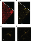
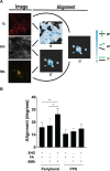
Methods: PPS cryosections from nonglaucomatous eyes were labelled for nuclei, fibrillar actin (FA), and alpha smooth muscle actin (αSMA) and imaged. Collagen fibrils were imaged using second harmonic generation. Nuclear density and aspect ratio of the internal PPS (iPPS), outer PPS (oPPS), and peripheral sclera were determined. FA and αSMA fibril alignment with collagen extracellular matrix (ECM) was determined. PPS fibroblasts were cultured on smooth or patterned membranes under mechanical strain and in the presence of TGFβ1 and 2.
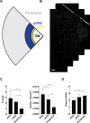
Results: The iPPS (7.1 ± 2.0 × 10⁻⁴, P < 0.0001) and oPPS (5.3 ± 1.4 × 10⁻⁴, P = 0.0013) had greater nuclei density (nuclei/µm2) than peripheral sclera (2.5 ± 0.8 × 10⁻⁴). The iPPS (2.0 ± 0.3, P = 0.002) but not oPPS (2.4 ± 0.4, P = 0.45) nuclei had smaller aspect ratios than peripheral (2.7 ± 0.5) nuclei. FA was present throughout the scleral stroma and was more aligned with oPPS collagen (9.6 ± 1.9 degrees) than in the peripheral sclera (15.9 ± 3.9 degrees, P =0.002). The αSMA fibers in the peripheral sclera were less aligned with collagen fibrils (26.4 ± 4.8 degrees) than were FA (15.9 ± 3.9 degrees, P = 0.0002). PPS fibroblasts cultured on smooth membranes shifted to an orientation perpendicular to the direction of cyclic uniaxial strain (1 Hz, 5% strain, 42.2 ± 7.1 degrees versus 62.0 ± 8.5 degrees, P < 0.0001), whereas aligned fibroblasts on patterned membranes were resistant to strain-induced reorientation (5.9 ± 1.4 degrees versus 10 ± 3.3 degrees, P = 0.21). Resistance to re-orientation was reduced by TGFβ treatment (10 ± 3.3 degrees without TGFβ1 compared to 23.1 ± 4.5 degrees with TGFβ1, P < 0.0001).
Conclusions: Regions of the posterior sclera differ in cellular density and nuclear morphology. Topography alters the cellular response to mechanical strain.
Conflict of interest statement
Disclosure:
Figures


Similar articles
-
Analysis of 2-dimensional regional differences in the peripapillary scleral fibroblast cytoskeleton of normotensive and hypertensive mouse eyes.Sci Rep. 2025 Jul 1;15(1):21207. doi: 10.1038/s41598-025-08251-4. Sci Rep. 2025. PMID: 40595239 Free PMC article.
-
Cyclic strain alters the transcriptional and migratory response of scleral fibroblasts to TGFβ.Exp Eye Res. 2024 Jul;244:109917. doi: 10.1016/j.exer.2024.109917. Epub 2024 Apr 30. Exp Eye Res. 2024. PMID: 38697276
-
Peripapillary sclera architecture revisited: A tangential fiber model and its biomechanical implications.Acta Biomater. 2018 Oct 1;79:113-122. doi: 10.1016/j.actbio.2018.08.020. Epub 2018 Aug 21. Acta Biomater. 2018. PMID: 30142444 Free PMC article.
-
Dasatinib inhibits peripapillary scleral myofibroblast differentiation.Exp Eye Res. 2020 May;194:107999. doi: 10.1016/j.exer.2020.107999. Epub 2020 Mar 13. Exp Eye Res. 2020. PMID: 32179077 Free PMC article.
-
High-Magnitude and/or High-Frequency Mechanical Strain Promotes Peripapillary Scleral Myofibroblast Differentiation.Invest Ophthalmol Vis Sci. 2015 Dec;56(13):7821-30. doi: 10.1167/iovs.15-17848. Invest Ophthalmol Vis Sci. 2015. PMID: 26658503 Free PMC article.
Cited by
-
Transcriptomic analysis of the ocular posterior segment completes a cell atlas of the human eye.Proc Natl Acad Sci U S A. 2023 Aug 22;120(34):e2306153120. doi: 10.1073/pnas.2306153120. Epub 2023 Aug 11. Proc Natl Acad Sci U S A. 2023. PMID: 37566633 Free PMC article.
-
Ocular Biomechanics and Glaucoma.Vision (Basel). 2023 Apr 23;7(2):36. doi: 10.3390/vision7020036. Vision (Basel). 2023. PMID: 37218954 Free PMC article. Review.
-
Transcriptomic Analysis of the Ocular Posterior Segment Completes a Cell Atlas of the Human Eye.bioRxiv [Preprint]. 2023 Apr 27:2023.04.26.538447. doi: 10.1101/2023.04.26.538447. bioRxiv. 2023. Update in: Proc Natl Acad Sci U S A. 2023 Aug 22;120(34):e2306153120. doi: 10.1073/pnas.2306153120. PMID: 37162855 Free PMC article. Updated. Preprint.
-
Analysis of 2-dimensional regional differences in the peripapillary scleral fibroblast cytoskeleton of normotensive and hypertensive mouse eyes.Sci Rep. 2025 Jul 1;15(1):21207. doi: 10.1038/s41598-025-08251-4. Sci Rep. 2025. PMID: 40595239 Free PMC article.
-
Combinatorial extracellular matrix cues with mechanical strain induce differential effects on myogenesis in vitro.Biomater Sci. 2023 Aug 22;11(17):5893-5907. doi: 10.1039/d3bm00448a. Biomater Sci. 2023. PMID: 37477446 Free PMC article.
References
-
- Quigley HA, Addicks EM, Green WR, Maumenee AE.. Optic nerve damage in human glaucoma. II. The site of injury and susceptibility to damage. Arch Ophthalmol. 1981; 99(4): 635–649. - PubMed
-
- Sigal IA, Flanagan JG, Ethier CR.. Factors influencing optic nerve head biomechanics. Invest Ophthalmol Vis Sci. 2005; 46(11): 4189–4199. - PubMed
Publication types
MeSH terms
Substances
Grants and funding
LinkOut - more resources
Full Text Sources
Other Literature Sources
Miscellaneous

