. 2021 Jan 4;62(1):28.
doi: 10.1167/iovs.62.1.28.

Ambient Light Regulates Retinal Dopamine Signaling and Myopia Susceptibility

Affiliations

Ambient Light Regulates Retinal Dopamine Signaling and Myopia Susceptibility

Erica G Landis et al. Invest Ophthalmol Vis Sci. .

Abstract

Purpose: Exposure to high-intensity or outdoor lighting has been shown to decrease the severity of myopia in both human epidemiological studies and animal models. Currently, it is not fully understood how light interacts with visual signaling to impact myopia. Previous work performed in the mouse retina has demonstrated that functional rod photoreceptors are needed to develop experimentally-induced myopia, alluding to an essential role for rod signaling in refractive development.

Methods: To determine whether dim rod-dominated illuminance levels influence myopia susceptibility, we housed male C57BL/6J mice under 12:12 light/dark cycles with scotopic (1.6 × 10⁻³ candela/m2), mesopic (1.6 × 101 cd/m2), or photopic (4.7 × 10³ cd/m2) lighting from post-natal day 23 (P23) to P38. Half the mice received monocular exposure to -10 diopter (D) lens defocus from P28-38. Molecular assays to measure expression and content of DA-related genes and protein were conducted to determine how illuminance and lens defocus alter dopamine (DA) synthesis, storage, uptake, and degradation and affect myopia susceptibility in mice.

Results: We found that mice exposed to either scotopic or photopic lighting developed significantly less severe myopic refractive shifts (lens treated eye minus contralateral eye; -1.62 ± 0.37D and -1.74 ± 0.44D, respectively) than mice exposed to mesopic lighting (-3.61 ± 0.50D; P < 0.005). The 3,4-dihydroxyphenylacetic acid /DA ratio, indicating DA activity, was highest under photopic light regardless of lens defocus treatment (controls: 0.09 ± 0.011 pg/mg, lens defocus: 0.08 ± 0.008 pg/mg).

Conclusions: Lens defocus interacted with ambient conditions to differentially alter myopia susceptibility and DA-related genes and proteins. Collectively, these results show that scotopic and photopic lighting protect against lens-induced myopia, potentially indicating that a broad range of light levels are important in refractive development.


Conflict of interest statement

Disclosure: E.G. Landis, None; H.N. Park, None; M. Chrenek, None; L. He, None; C. Sidhu, None; R. Chakraborty, None; R. Strickland, None; P.M. Iuvone, None; M.T. Pardue, None

Figures

Figure 1.
Figure 1.
Experimental Design of the Study.
Figure 2.
Figure 2.
Scotopic and photopic light exposure inhibited lens induced myopia in mice. (A) Control mice housed in scotopic (black), mesopic (blue), or photopic (orange) light showed identical refractive error development. (B) The myopic shift at P35 of lens treated mice exposed to mesopic light is greater than mice exposed to scotopic or photopic light (one-way ANOVA F(2,58) = 6.50; P < 0.01). All data shown are mean ± SEM, n = 17–21/lens defocus group, post hoc comparisons indicated by **P < 0.01.
Figure 3.
Figure 3.
Axial length correlated with refractive error in control and mesopic housed lens defocus mice. (A) In control animals, axial length and refractive error at 28 and 35 days of age were positively correlated under scotopic (black circles, R2 = 0.407; P < 0.001), mesopic (blue squares, R2 = 0.368; P < 0.01) and photopic (orange triangles, R2 = 0.249; P < 0.05) light. (B) With lens defocus treatment, mice housed in either scotopic or photopic light showed a nonsignificant, positive correlation between axial length and refractive error at 28 and 35 days for lens-treated eyes. However, mesopic light exposure resulted in a negative correlation between refractive error and axial length (R2 = 0.261; P < 0.05), indicating more myopic refractive errors were associated with longer axial lengths. n = number of eyes used in the analysis at both timepoints; only right eyes are shown for both panels.
Figure 4.
Figure 4.
Lens treatment with mesopic light exposure steepened corneal curvature. (A) Corneal curvature of mice housed in scotopic light did not change with lens treatment but did increase with age (RM two-way ANOVA, main effect of age, F(2, 108) = 234.8; P < 0.001). (B) In mesopic light, the lens-treated eye had the lowest corneal curvature at P35 (RM two-way ANOVA, interaction effect, F(4, 108) = 3.7; P < 0.01). (C) Corneal curvature of mice housed in photopic light increased with age (RM two-way ANOVA, main effect of age, F(2, 106) = 195.6; P < 0.001) but did not change with lens treatment. Control eyes are shown in solid black lines, the naïve left eyes are shown in red dashed lines, and lens-treated eyes are shown in solid red lines. All data shown is mean ± SEM.
Figure 5.
Figure 5.
DA turnover increases with illuminance level. Retinas were collected four to six hours after light onset under each housing illumination level. (A) After ∼2 weeks of ambient light exposure, DOPAC levels were increased with higher light intensities in both treatment groups (two-way ANOVA, main effect of light, F(2,53) = 18.47; P < 0.001). For both treatment groups, all light level groups had significantly different DOPAC levels (P < 0.01). (B) None of the treatment groups showed changes in DA levels with light or lens defocus. (C) DOPAC/DA ratio increased with light indicating higher dopamine metabolism at higher light intensities (two-way ANOVA, main effect of light, F(2,53) = 19.45; P < 0.001). For both treatment groups, all light level groups had significantly different DOPAC/DA ratios (P < 0.05). Scotopic samples are represented by black circles, mesopic samples by blue squares, and photopic samples by orange triangles. Bars represent mean ± SEM.
Figure 6.
Figure 6.
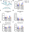
Expression of genes associated with DA signaling. The expression levels of DA signaling genes were measured with ddPCR after light exposure and LIM. (A) DA signaling in dopaminergic cells depend on tyrosine hydroxylase and several other related proteins to tightly control levels of DA and its metabolite DOPAC. (B) In control mice, Th expression was significantly higher in mice housed in photopic light compared to scotopic (two-way ANOVA, interaction effect F(2,29) = 7.52; P < 0.01; post hoc comparison; P < 0.05). Lens defocus–treated retinas from mesopic light had significantly higher Th expression then lens treated, photopic retinas (P < 0.05). (C, D) No significant differences were found in expression of Slc6a3 (DAT) or Slc18a2 (VMAT2). (E) LIM eyes were significantly higher than control eyes for expression levels of Maoa (two-way ANOVA, main effect of treatment, F(1,29) = 9.92; P < 0.01) and (F) Maob expression did not change with lens or light treatments . Data are mean ± SEM measured in arbitrary units normalized to levels of HPRT. Scotopic samples are represented by black circles, mesopic samples by blue squares, and photopic samples by orange triangles. For post hoc comparisons, *P < 0.05.
Figure 7.
Figure 7.
Decreased phosphorylated TH under lens treatment as ambient light increases. (A) Levels of TH did not change significantly with ambient light or LIM. (B) Phosphorylated TH, pTHSer40, was inversely related to light intensity (two-way ANOVA, main effect of treatment, F(1,33) = 8.34; P < 0.01). This effect was likely driven by decreased pTHSer40 levels in lens defocus–treated retinas housed in photopic light (orange triangles) compared to mesopic lens–treated retinas (blue squares) and scotopic lens–treated retinas (black circles) compared to equivalent pTHSer40 in control retinas from all light levels. (C) The ratio of pTHSer40 to total TH showed a nonsignificant relationship between control and lens defocused retinas under each light level. Only retinas which were used for TH and pTHSer40 labeling were used in this analysis limiting the sample number. (D) VMAT2 protein levels were not significantly affected by light or lens defocus exposure. Data are mean ± SEM. Representative blots are included in Supplementary Figure S4.
Figure 8.
Figure 8.
Acute exposure to photopic illuminance drives higher DA activity. (A) The levels of DOPAC in retinas exposed to three hours of scotopic (black circles, n = 5–7), mesopic (blue squares, n = 5–10), or photopic (orange triangles, n = 5–9) light indicate an increase in DA metabolism under bright light during short exposures (one-way ANOVA, F(2, 23) = 27.59; P < 0.001). (B) DA was highest in scotopic light (one-way ANOVA, F(2, 23) = 7.32; P < 0.01). (C) The DOPAC/DA ratio representing DA activity was lowest in scotopic exposed retinas and highest in photopic light (one-way ANOVA, F(2, 23) = 35.22; P < 0.001). The gene expression of (D) Th, (E) Slc18a2, (F) Slc6a3 were not significantly different across illuminance level exposures. Data represent as mean ± SEM. Post hoc comparisons are indicated, **P < 0.01, ***P < 0.001.
Figure 9.
Figure 9.
Light sensitivity across photoreceptors. The human photoreceptor sensitivity covers a broad range of natural and artificial light. Scotopic light is below the cone threshold of activation, mesopic light activates both rod and cone photoreceptors, and photopic light activates cone photoreceptors. Recent research has indicated potential roles for melanopsin retinal ganglion cells (mRGCs) and rods in photopic light., The illuminance levels used in these experiments are indicated with black arrows. Figure is adapted from references and to .

Similar articles

Cited by

References

    1. Vitale S, Sperduto RD, Ferris FL 3rd. Increased prevalence of myopia in the United States between 1971-1972 and 1999-2004. Arch Ophthalmol. 2009; 127: 1632–1639. - PubMed
    1. Li J, Zhang Q.. Insight into the molecular genetics of myopia. Mol Vis. 2017; 23: 1048–1080. - PMC - PubMed
    1. Troilo D, Smith EL 3rd, Nickla DL, et al. .. IMI - Report on Experimental Models of Emmetropization and Myopia. Invest Ophthalmol Vis Sci. 2019; 60: M31–M88. - PMC - PubMed
    1. Wildsoet CF, Chia A, Cho P, et al. .. IMI - Interventions Myopia Institute: Interventions for Controlling Myopia Onset and Progression Report. Invest Ophthalmol Vis Sci. 2019; 60: M106–M131. - PubMed
    1. Mutti DO, Mitchell GL, Moeschberger ML, Jones LA, Zadnik K.. Parental myopia, near work, school achievement, and children's refractive error. Invest Ophthalmol Vis Sci. 2002; 43: 3633–3640. - PubMed

Publication types

MeSH terms

Substances