. 2021 Apr 1;162(4):bqab015.
doi: 10.1210/endocr/bqab015.

PKA and AMPK Signaling Pathways Differentially Regulate Luteal Steroidogenesis

Affiliations

PKA and AMPK Signaling Pathways Differentially Regulate Luteal Steroidogenesis

Emilia Przygrodzka et al. Endocrinology. .

Abstract

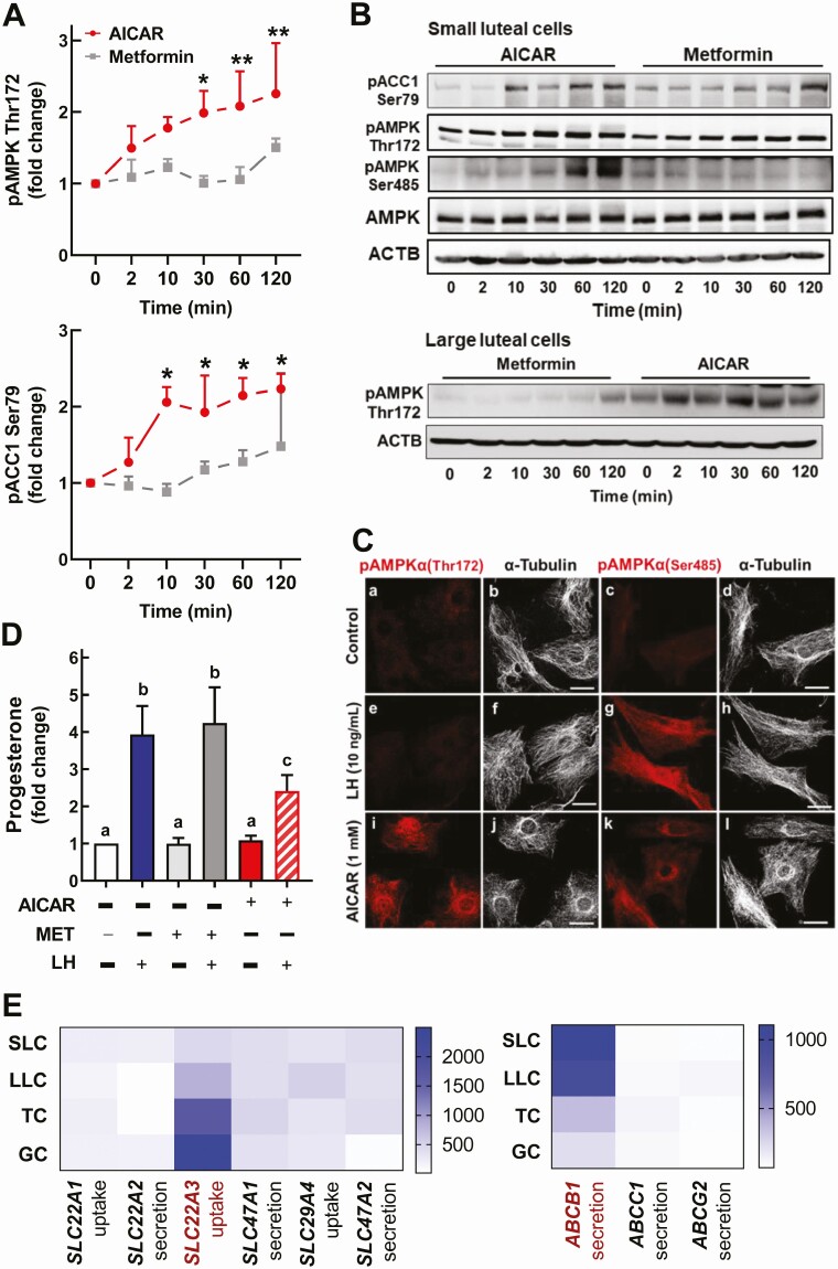
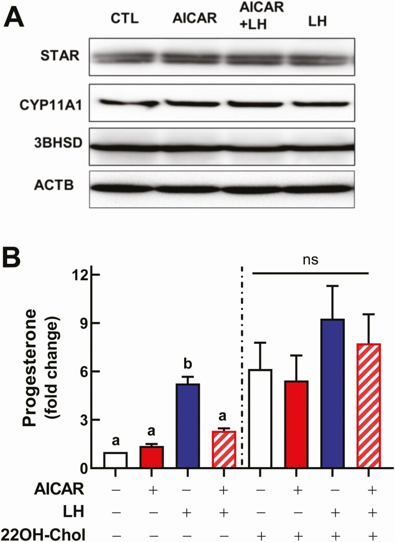
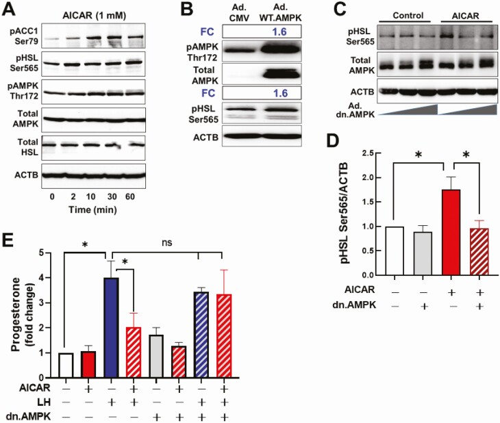
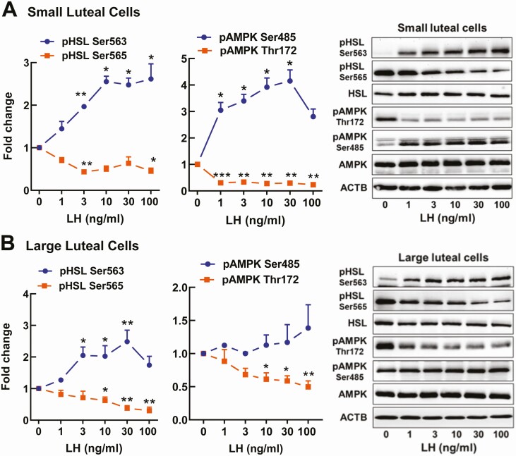
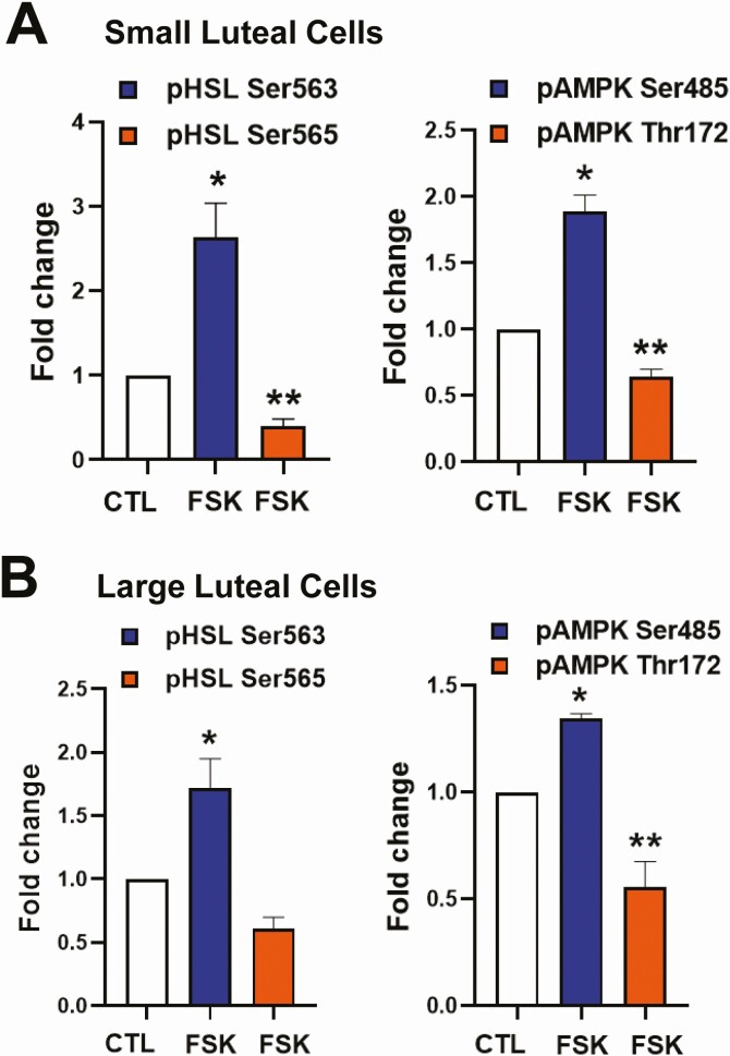
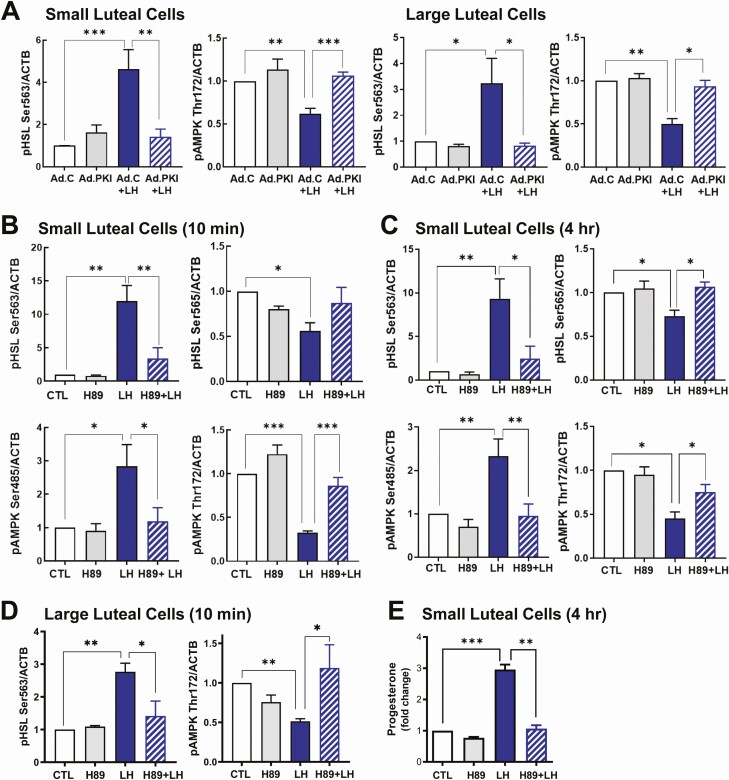
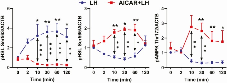
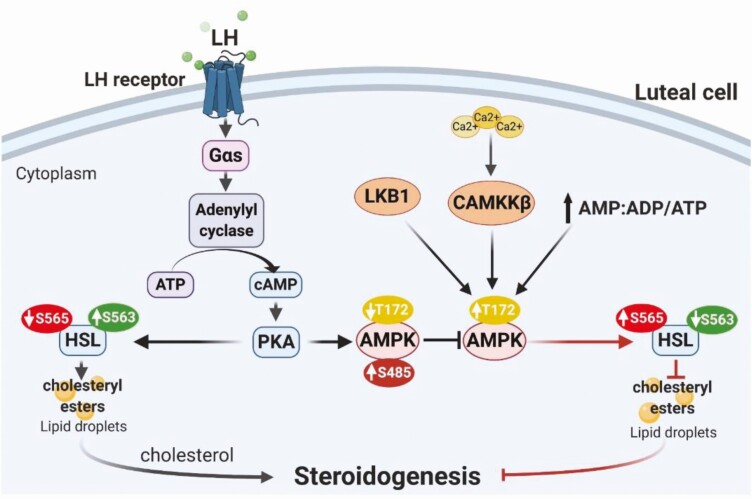
Luteinizing hormone (LH) via protein kinase A (PKA) triggers ovulation and formation of the corpus luteum, which arises from the differentiation of follicular granulosa and theca cells into large and small luteal cells, respectively. The small and large luteal cells produce progesterone, a steroid hormone required for establishment and maintenance of pregnancy. We recently reported on the importance of hormone-sensitive lipase (HSL, also known as LIPE) and lipid droplets for appropriate secretory function of the corpus luteum. These lipid-rich intracellular organelles store cholesteryl esters, which can be hydrolyzed by HSL to provide cholesterol, the main substrate necessary for progesterone synthesis. In the present study, we analyzed dynamic posttranslational modifications of HSL mediated by PKA and AMP-activated protein kinase (AMPK) as well as their effects on steroidogenesis in luteal cells. Our results revealed that AMPK acutely inhibits the stimulatory effects of LH/PKA on progesterone production without reducing levels of STAR, CYP11A1, and HSD3B proteins. Exogenous cholesterol reversed the negative effects of AMPK on LH-stimulated steroidogenesis, suggesting that AMPK regulates cholesterol availability in luteal cells. AMPK evoked inhibitory phosphorylation of HSL (Ser565). In contrast, LH/PKA decreased phosphorylation of AMPK at Thr172, a residue required for its activation. Additionally, LH/PKA increased phosphorylation of HSL at Ser563, which is crucial for enzyme activation, and decreased inhibitory phosphorylation of HSL at Ser565. The findings indicate that LH and AMPK exert opposite posttranslational modifications of HSL, presumptively regulating cholesterol availability for steroidogenesis.

Keywords: corpus luteum; hormone-sensitive lipase; lipid droplets; luteinizing hormone; progesterone; protein kinase.


Figures

Figure 1.
Figure 1.
The AMPK activator AICAR, but not metformin, inhibits LH-stimulated production of progesterone by luteal cells. (A-B) Primary cultures of bovine small and large luteal cells were treated with the AMPK activators, AICAR (1mM; ●   ) or Metformin (25mM; ■) for 2 to 120 minutes. Line graphs (A) and representative immunoblots (B) showing phosphorylation of AMPKα Thr172, AMPKα Ser485, and the AMPK substrate ACC1 Ser79. (C) Representative confocal micrographs of phosphorylated AMPKα Thr172 and Ser485 in primary bovine small luteal cells following treatment with control media (panels a-d), LH (10 ng/mL) for 30 minutes (panels e-h), or AICAR (1mM) for 1 hour (panels i-l). (D) Small luteal cell cultures were pretreated with AICAR (1mM) or metformin (25mM) for 1 hour and then treated with LH (10 ng/mL) for 4 hours. Progesterone in conditioned media was measured by ELISA. Data are shown as a fold change and presented as means ± SEM from 4 to 7 separate experiments. Statistical analysis was performed using 1-way ANOVA test with post hoc Bonferroni test. (E) Heatmaps showing expression of metformin transporters in small luteal cells (SLC), large luteal cells (LLC), granulosa cells (GC), and theca cells (TC). Data were obtained from previous studies (55). Abbreviations: SLC22A1-3 (Solute Carrier Family 22 Member 1-3); SLC29A4 (Solute Carrier Family 29 Member 4; SLC47A1 (Solute Carrier Family 47 Member 1); ABCB1 (ATP Binding Cassette Subfamily B Member 1); ABCC1 (ATP Binding Cassette Subfamily C Member 1); ABCG2 (ATP Binding Cassette Subfamily G Member 2). Panel A: Data were normalized to β-actin (ACTB). Significant differences from zero time control are indicated with asterisks; * and ** represent P < 0.05 and P < 0.01, respectively. Panel D: Bars with different lettersabc are significantly different; P < 0.05.
Figure 2.
Figure 2.
Exogenous cholesterol reverses inhibitory effects of AMPK on LH-stimulated production of progesterone. Panel A: Representative blots showing content of STAR, CYP11A1, and HSD3B in the primary small luteal cells pretreated with AICAR (1mM) for 1 hour and then treated with LH (10 ng/mL) for 4 hours. Panel B: Bovine luteal cells were pretreated with AICAR (1mM) for 1 hour and then treated with or without 22-hydoxycholesterol (22-OH Chol; 15 μM) in the presence or absence of LH (10 ng/mL) for 4 hours. Progesterone measured by ELISA. Data are shown as a fold change and presented as means ± SEM from 4 separate experiments. Statistical analysis was performed using 1-way ANOVA test with post hoc Bonferroni test. Bars with different lettersabc are significantly different; P < 0.05.
Figure 3.
Figure 3.
AMPK affects posttranslational modification of enzyme involved in lipolysis. (A) Small luteal cells were treated with AICAR (1mM) for 2, 10, 30, and 60 minutes. Representative Western blots of phosphorylation of AMPKα and downstream targets HSL Ser565 and ACC1 Ser79. Total AMPK, HSL, and β-actin (ACTB) are shown. (B) Small luteal cells were infected with control adenovirus Ad.CMV or adenovirus expressing wild-type AMPKα (Ad.WT.AMPK). Representative blots showing levels of phospho-AMPKα (Thr172), phospho-HSL (Ser565), and total AMPKα. (C-D) Small luteal cells were infected with control adenovirus (Ad.CMV) or adenovirus expressing dominant negative AMPKα1 (Ad.dn.AMPK) for 48 hours and then treated with AICAR (1mM) for 15 minutes. Representative blots (Panel C) and bar graph (Panel D) showing levels of phospho-HSL Ser565 and total AMPKα. Significant differences between treatments are indicated with asterisks * reflecting P < 0.05, n = 4. (E) Small luteal cells were infected with Ad.dn.AMPK and then pretreated with AICAR (1mM) for 1 hour followed by incubation with LH (10 ng/mL) for 4 hours. Progesterone was measured by ELISA. Panels D and E: Data are shown as a fold change and presented as means ± SEM from 3 to 4 separate experiments. Statistical analysis was performed using 1-way ANOVA test with post hoc Bonferroni test. * P < 0.05.
Figure 4.
Figure 4.
LH affects phosphorylation of AMPKα and HSL. Primary cultures of small luteal cells (A) and large luteal cells (B) were treated with increasing concentrations of LH (0-100 ng/mL) for 30 minutes. Line graphs and representative blots showing phosphorylation of HSL (Ser563 and 565) and AMPKα (Thr172 and Ser485). Panels A and B: Data are shown as the average fold changes ± SEM from 3 to 5 separate experiments. Data were normalized to β-actin (ACTB). Statistical analysis was performed using 1-way ANOVA with post hoc Bonferroni tests. Significant differences between LH concentrations and control are indicated with asterisks *, **, *** reflecting P < 0.05, P < 0.01, P < 0.001, respectively.
Figure 5.
Figure 5.
Activation of the adenylyl cyclase/PKA pathway mimics the action of LH on AMPKα and HSL phosphorylation. Cultures of small (A) and large luteal cells (B) were treated with control medium (CTL) or forskolin (FSK; 10µM) for 30 minutes. Western blot analysis was performed to evaluate phosphorylation of HSL (Ser563 and Ser565) and AMPKα (Thr1712 and Ser485). Data were normalized to β-actin. Bar graphs show average fold changes ± SEM from 3-5 separate experiments. Statistical analysis was performed using 1-way ANOVA test for repeated measurements and Bonferroni post hoc test. Significant differences are indicated with asterisks * and ** representing P < 0.05 and P < 0.01, respectively.
Figure 6.
Figure 6.
The effect of LH on posttranslational modifications of HSL and AMPK is mediated by protein kinase A. (A) Primary cultures of small and large luteal cells were infected with a control adenovirus (Ad.C) or adenovirus expressing the endogenous inhibitor of PKA (Ad.PKI). After 48 hours, cells were treated with LH (10 ng/mL) for 30 minutes. (B) Small luteal cells were pretreated with the PKA inhibitor H89 (30 μM) for 1 hour and then treated with LH (10 ng/mL) for 10 minutes. (C) Small luteal cells were pretreated with H89 (30µM) for 1 hour and treated with LH (10 ng/mL) for 4 hours. (D) Large luteal cells were pretreated with the PKA inhibitor H89 (30 μM) for 1 hour and then treated with LH (10 ng/mL) for 10 minutes. (E) Small luteal cells were pretreated with H89 (30µM) for 1 hour and treated with LH (10 ng/mL) for 4 hours. Media samples were collected for analysis of progesterone using ELISA. Bar graphs represent average fold changes ± SEM from 3-5 separate experiments. Statistical analysis was done using 1-way ANOVA with Bonferroni post hoc test. Significant differences between groups are indicated with asterisks *, **, *** reflecting P < 0.05, P < 0.01, P < 0.001, respectively.
Figure 7.
Figure 7.
AICAR suppresses the effects of LH on posttranslational modifications of HSL and AMPK. Cultures of small luteal cells were pretreated without (●   ) or with AICAR (1mM, ■) for 1 hour and then treated with LH (10 ng/mL) for up to 120 minutes. Line graphs showing phosphorylation of HSL Ser563, HSL 565, and AMPKα Thr172 are shown as average fold changes ± SEM from 3 to 5 separate experiments. Statistical analysis was performed using 1-way (LH versus control) or 2-way ANOVA (↔, LH versus LH + AICAR) with post hoc Bonferroni test. Significant differences are indicated with asterisks *, **, and *** as P < 0.05, P < 0.01 and P < 0.001, respectively.
Figure 8.
Figure 8.
Proposed model of LH action in small and large luteal cells. LH binds to G-protein coupled receptors present on the surface of luteal cells leading to increased activity of protein kinase A (PKA). LH/PKA increases the activity of HSL by triggering phosphorylation of HSL at Ser563 (S563) and reducing phosphorylation at Ser565 (S565). Active HSL translocates to lipid droplets and cleaves cholesteryl esters to release cholesterol for progesterone production by luteal cells. Simultaneously, LH reduces phosphorylation of AMPKα at Thr172 and phosphorylates AMPKα at Ser485 (S485) preventing its phosphorylation at Thr172 (T172) by upstream kinases, ie, calcium/calmodulin-dependent protein kinase kinase 2 (CAMKKβ), liver kinase b1 (LKB1), or increased AMP/ATP ratio. In case of luteolysis, upstream kinases (CAMKK, LKB1) or increased AMP: ATP ratio activate AMPK by triggering its phosphorylation at Thr172. Active AMPK phosphorylates HSL at Ser565, which inhibits phosphorylation at Ser563 leading to inactivation of HSL and preventing hydrolysis of cholesteryl esters stored in lipid droplets and limiting cholesterol availability. Consequently, the elevated activity of AMPK inhibits steroidogenic capacity of luteal cells.

Similar articles

Cited by

References

    1. Berisha B, Schams D. Ovarian function in ruminants. Domest Anim Endocrinol. 2005;29(2):305-317. - PubMed
    1. Diskin MG, Morris DG. Embryonic and early foetal losses in cattle and other ruminants. Reprod Domest Anim. 2008;43(Suppl 2):260-267. - PubMed
    1. Lonergan P. Influence of progesterone on oocyte quality and embryo development in cows. Theriogenology. 2011;76(9):1594-1601. - PubMed
    1. Davis JS, Rueda BR. The corpus luteum: an ovarian structure with maternal instincts and suicidal tendencies. Front Biosci. 2002;7:d1949-d1978. - PubMed
    1. McCracken JA, Carlson JC, Glew ME, et al. Prostaglandin F 2 identified as a luteolytic hormone in sheep. Nat New Biol. 1972;238(83):129-134. - PubMed

Publication types