. 2021 Mar 3;223(5):752-764.
doi: 10.1093/infdis/jiab039.

Evidence of Severe Acute Respiratory Syndrome Coronavirus 2 Replication and Tropism in the Lungs, Airways, and Vascular Endothelium of Patients With Fatal Coronavirus Disease 2019: An Autopsy Case Series

Affiliations

Evidence of Severe Acute Respiratory Syndrome Coronavirus 2 Replication and Tropism in the Lungs, Airways, and Vascular Endothelium of Patients With Fatal Coronavirus Disease 2019: An Autopsy Case Series

Julu Bhatnagar et al. J Infect Dis. .

Abstract

Background: The coronavirus disease 2019 (COVID-19) pandemic continues to produce substantial morbidity and mortality. To understand the reasons for the wide-spectrum complications and severe outcomes of COVID-19, we aimed to identify cellular targets of severe acute respiratory syndrome coronavirus 2 (SARS-CoV-2) tropism and replication in various tissues.

Methods: We evaluated RNA extracted from formalin-fixed, paraffin-embedded autopsy tissues from 64 case patients (age range, 1 month to 84 years; 21 COVID-19 confirmed, 43 suspected COVID-19) by SARS-CoV-2 reverse-transcription polymerase chain reaction (RT-PCR). For cellular localization of SARS-CoV-2 RNA and viral characterization, we performed in situ hybridization (ISH), subgenomic RNA RT-PCR, and whole-genome sequencing.

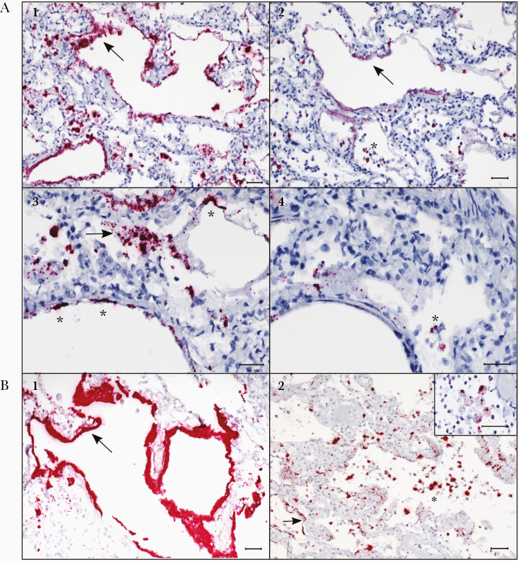
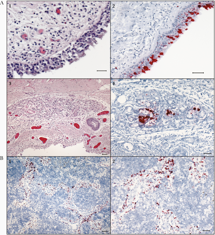
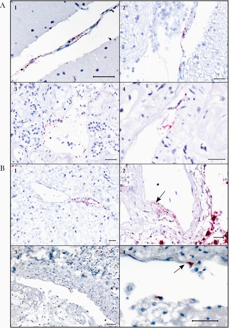
Results: SARS-CoV-2 was identified by RT-PCR in 32 case patients (21 COVID-19 confirmed, 11 suspected). ISH was positive in 20 and subgenomic RNA RT-PCR was positive in 17 of 32 RT-PCR-positive case patients. SARS-CoV-2 RNA was localized by ISH in hyaline membranes, pneumocytes, and macrophages of lungs; epithelial cells of airways; and endothelial cells and vessel walls of brain stem, leptomeninges, lung, heart, liver, kidney, and pancreas. The D614G variant was detected in 9 RT-PCR-positive case patients.

Conclusions: We identified cellular targets of SARS-CoV-2 tropism and replication in the lungs and airways and demonstrated its direct infection in vascular endothelium. This work provides important insights into COVID-19 pathogenesis and mechanisms of severe outcomes.

Keywords: COVID-19; SARS-CoV-2; autopsy; in situ hybridization; replication.


Figures

Figure 1.
Figure 1.
Localization of severe acute respiratory syndrome coronavirus 2 (SARS-CoV-2) RNA in hyaline membranes, pneumocytes, and intra-alveolar macrophages in the lungs of case patients with fatal coronavirus disease 2019 (COVID-19) by in situ hybridization (ISH) assays. A1, ISH (in red) targeting the N-gene of SARS-CoV-2 strongly stains hyaline membranes lining alveolar spaces (arrow) throughout the lung. Case patient 2. A2, ISH targeting the S-gene in a serial section from the same case as image A1, showing staining in hyaline membranes (arrow), and highlighting staining in intra-alveolar cells consistent with sloughed pneumocytes and alveolar macrophages (asterisk). Case patient 2. A3, Higher magnification image showing N-gene ISH staining in hyaline membranes (arrow) and pneumocytes (asterisks) lining alveolar spaces in the lung. Case patient 2. A4, Serial section from the same case as image A3, illustrating staining by ISH targeting the S-gene, showing staining in hyaline membranes and scattered staining in intra-alveolar cells (asterisk). Case patient 2. B1, Abundant staining by the N-gene ISH assay in hyaline membranes (arrow) in the lung of an acute case of COVID-19. Case patient 30. B2. Stippled staining by the N-gene ISH assay in hyaline membranes (arrow) and strong staining in intra-alveolar cells (asterisk), consistent with sloughed pneumocytes and intra-alveolar macrophages (macrophages highlighted in inset), in the lungs of COVID-19 autopsy cases. Case patients 11 and 23 (inset). All scale markers represent 50 µM.
Figure 2.
Figure 2.
In situ hybridization (ISH) assays demonstrating severe acute respiratory syndrome coronavirus 2 (SARS-CoV-2) RNA in the tracheal epithelium and tracheal submucosal glands, and in macrophages within lymph node of patients with fatal coronavirus disease 2019 (COVID-19). A1, Section of trachea showing moderate mononuclear infiltrates in the submucosa of the trachea. Hematoxylin and eosin (H&E) stain. Case patient 12. A2, Multifocal, regional ISH staining of respiratory epithelium within the trachea. ISH for S-gene of SARS-CoV-2. Case patient 12. A3, Photomicrograph of a submucosal gland within the trachea showing mild, mononuclear infiltrates within and around the glands, H&E. Case patient 13. A4, ISH staining within the glandular epithelium of the submucosal gland. ISH for N-gene of SARS-CoV-2. Case patient 13. B1, ISH staining for the N-gene of SARS-CoV-2 within macrophages in the medullary sinuses of the mediastinal lymph node. Case patient 17. B2, Higher-magnification image of ISH staining in macrophages within medullary sinuses of the mediastinal lymph node. Case patient 17. All scale markers represent 50 µM.
Figure 3.
Figure 3.
Endothelial and vascular wall staining in multiple tissues by N-gene in situ hybridization (ISH) assay in a fatal case of coronavirus disease 2019 (COVID-19) (case patient 30). Also note ISH staining in endothelial cells associated with a thrombus in another COVID-19 fatal case (case patient 22). A1, Stippled ISH staining (red) in the endothelium and tunica media of a vessel in the cerebellar leptomeninges in a confirmed case of acute COVID-19. A2, Regional staining by ISH in the vascular endothelium and tunica media in the brain stem parenchyma. A3, Endothelial ISH staining within vessels in the kidney. A4, Stippled ISH staining in detached endothelial cells of a renal vessel. B1, Stippled ISH staining seen in the endothelium and tunica media of a vessel within the autolyzed pancreas. B2, ISH staining within the tunica media of a vessel in the lung (arrow); ISH staining of pneumocytes in the adjacent alveoli is also noted. B3, ISH staining in an endothelial cell and a circulating cell in a thrombosed vessel of an additional confirmed case of COVID-19. Thrombus indicated by an asterisk. B4, Higher-magnification image of the distinct endothelial cell staining from image B3 (arrow). All scale markers represent 50 µM.

Similar articles

Cited by

References

    1. Zhu N, Zhang D, Wang W, et al. A novel coronavirus from patients with pneumonia in China, 2019. N Engl J Med 2020; 382:727–33. - PMC - PubMed
    1. World Health Organization. Coronavirus disease (COVID-19). https://www.who.int/emergencies/diseases/novel-coronavirus-2019. Accessed 4 January 2021.
    1. Centers for Disease Control and Prevention. Coronavirus disease 2019 (COVID-19) in the U.S. https://covid.cdc.gov/covid-data-tracker/#cases_casesper100klast7days. Accessed 4 January 2021.
    1. Cevik M, Bamford CGG, Ho A. COVID-19 pandemic—a focused review for clinicians. Clin Microbiol Infect 2020; 26:842–7. - PMC - PubMed
    1. Solomon IH, Normandin E, Bhattacharyya S, et al. Neuropathological features of Covid-19. N Engl J Med 2020; 383:989–92. - PMC - PubMed

Publication types

MeSH terms