. 2021 May;384(2):499-511.
doi: 10.1007/s00441-020-03398-0. Epub 2021 Jan 27.

Identification of CXCL11 as part of chemokine network controlling skeletal muscle development

Affiliations

Identification of CXCL11 as part of chemokine network controlling skeletal muscle development

Malte Puchert et al. Cell Tissue Res. 2021 May.

Abstract

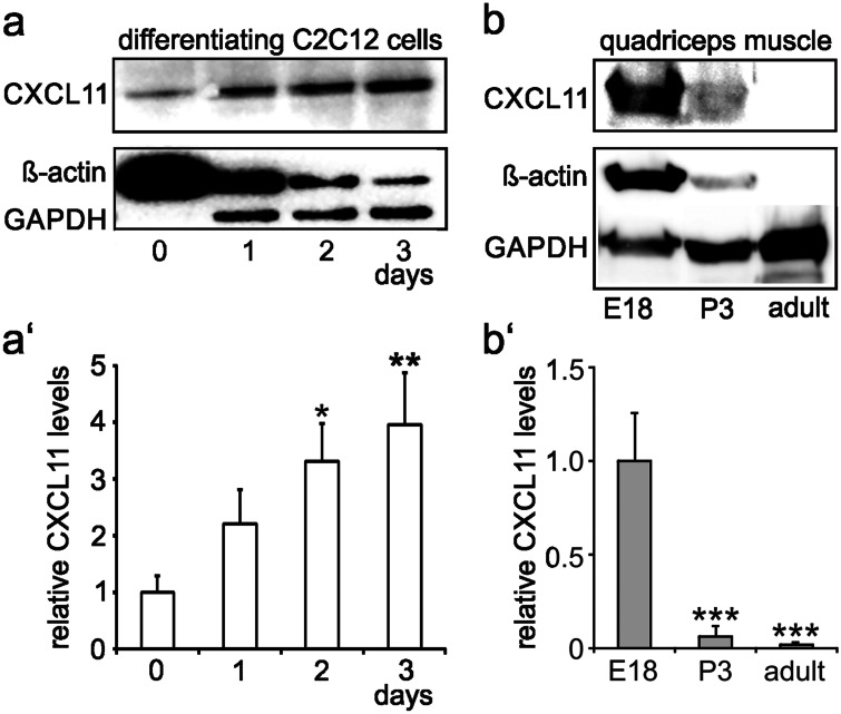
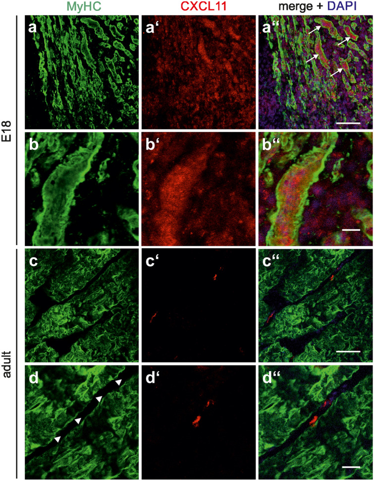
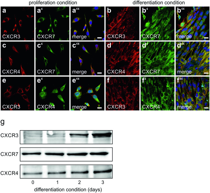
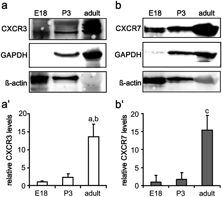
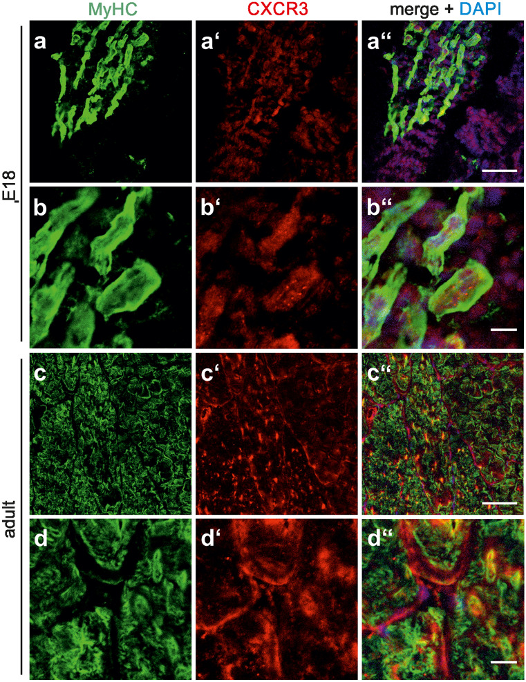
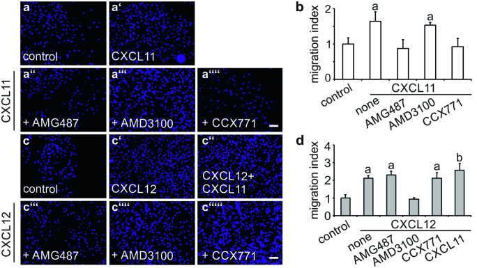
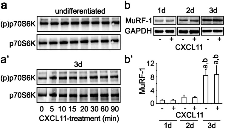
The chemokine, CXCL12, and its receptors, CXCR4 and CXCR7, play pivotal roles during development and maintenance of limb muscles. CXCR7 additionally binds CXCL11, which uses CXCR3 as its prime receptor. Based on this cross-talk, we investigate whether CXCL11 would likewise affect development and/or function of skeletal muscles. Western blotting and immunolabelling demonstrated the developmentally restricted expression of CXCL11 in rat limb muscles, which was contrasted by the continuous expression of its receptors in proliferating and differentiating C2C12 cells as well as in late embryonic to adult rat limb muscle fibres. Consistent with a prime role in muscle formation, functional studies identified CXCL11 as a potent chemoattractant for undifferentiated C2C12 cells and further showed that CXCL11 does neither affect myoblast proliferation and differentiation nor metabolic/catabolic pathways in formed myotubes. The use of selective receptor antagonists unravelled complementary effects of CXCL11 and CXCL12 on C2C12 cell migration, which either require CXCR3/CXCR7 or CXCR4, respectively. Our findings provide new insights into the chemokine network controlling skeletal muscle development and function and, thus, might provide a base for future therapies of muscular diseases.

Keywords: CXCL11; CXCR3; CXCR7; Myoblasts; Skeletal muscle fibres.


Conflict of interest statement

The authors declare that they have no conflict of interest.

Figures

Fig. 1
Fig. 1
Expression of CXCL11 during myogenesis. a Western blot analysis demonstrated that CXCL11 levels gradually increase in C2C12 cells following the switch to differentiation conditions (DMEM + 1% FCS). a′ Relative levels of CXCL11 (mean ± SD, n = 3) as determined by densitometric analysis of immunoreactive protein bands in C2C12 cells maintained under differentiation conditions for the indicated times. CXCL11 levels present prior to the switch to differentiation conditions (day 0) were set to 1. *p < 0.05, **p < 0.005, respective time point vs. day 0. b CXCL11 expression is readily detectable by Western blot analysis in the rat quadriceps muscle at E18 with a clear decline into adulthood. b′ relative levels of CXCL11 (mean ± SD) as determined by densitometry of immunoreactive protein bands from 3 independent experiments. ***p < 0.001, respective time point vs. E18. Note in a and b that ß-actin as well as GAPDH, which are routinely used as loading controls for Western blotting, are developmentally regulated in muscle cells/fibres. Due to this fact, all samples were carefully adjusted to identical protein levels prior to analysis
Fig. 2
Fig. 2
Localization of CXCL11 expression in developing and adult limb muscles. Sections were obtained from the quadriceps muscle of E18 a, b and adult rats c, d and double-labelled with antibodies against CXCL11 (red) and MyHC (green). Cell nuclei were visualized with DAPI. At each developmental stage, first row a, c and second row b, d show low and high magnification, respectively. a″d″ Merging of immunocytochemical stainings and DAPI. In E18 muscles, CXCL11-immunolabelling was associated with many muscle fibres (arrows in a″). Higher magnification b″ confirmed the predominant intracellular localization of CXCL11 at this developmental stage. In adult muscles, CXCL11 expression was confined to cells within the endomysium/perimysium (arrow heads in d). Scale bars, a and c, 100 µm; b and d, 25 µm
Fig. 3
Fig. 3
Expression and co-localization of chemokine receptors in C2C12 cells af C2C12 cells were maintained under either proliferation conditions a, c, e or differentiation conditions b, d, f for 3 days and double-labelled with antibodies against CXCR3 (bs-2209R), CXCR7, or CXCR4 as indicated. a″f″ Merging of immunocytochemical stainings and DAPI. Virtually all non-differentiated C2C12 cells expressed CXCR3 a, e. Expression of CXCR3 persisted in forming myotubes b, f. In addition to CXCR3, again virtually all non-differentiated C2C12 cells as well as forming mytotubes co-expressed CXCR7 a″, b″ and CXCR4 e″, f″. Virtually all proliferating and differentiating cells further co-expressed CXCR4 and CXCR7 c″, d″. Scale bars, 10 µm. g Quantification of chemokine receptors expression in differentiating C2C12 cells. C2C12 cells were maintained for the indicated times with differentiation medium and analysed for CXCR3, CXCR7 and CXCR4 by Western blotting. Expression of the chemokine receptors increased with ongoing differentiation. Due to the lack of appropriate loading controls, care was taken to adjust samples to identical protein levels
Fig. 4
Fig. 4
Expression levels of CXCL11 receptors in developing and adult limb muscles a, b Western blotting allowed the detection of the CXCL11 receptors, a CXCR3 and b CXCR7, in the quadriceps muscle of embryonic to adult rats with a most prominent increase in expression levels between P3 and adulthood. a′, b′ Relative protein levels of a′ CXCR3 and b′ CXCR7 (mean ± SD, n = 3) as determined by densitometric analysis of immnoreactive protein bands. Protein levels present at E18 were set to 1. Note again that ß-actin and GAPDH, used as loading controls, are developmentally regulated (see Fig. 1). ap < 0.001, adult vs E18; bp < 0.01, adult vs. P3; cp < 0.001, adult vs. E18 or P3
Fig. 5
Fig. 5
Localization of CXCR3 in developing and adult limb muscles. ad'' Sections of the quadriceps muscle from E18 a, a', b, b' and adult rats c, c', d, d' were double-labelled with antibodies against CXCR3 (red; bs-2209R) and MyHC (green) as indicated. a″d″ Merging of immunocytochemical stainings and DAPI. At each developmental stage, first row a, c and second row b, d show low and high magnification, respectively. In the embryonic muscle, CXCR3 is primarily located within MyHC-positive muscle fibres b″, whereas in adult muscles CXCR3 is associated with both the sarcolemma and intracellular structures d″. Scale bars, a and c, 100 µm; b and d, 25 µm
Fig. 6
Fig. 6
Comparison of the chemotactic responses of C2C12 cells to CXCL11 and CXCL12 a, c Chemotactic responses of C2C12 cells to a CXCL11 (100 ng/ml) and c CXCL12 (100 ng/ml) were assessed in a modified Boyden chamber as described under Materials and Methods. Cells were treated with the CXCR3 antagonist, a″, c‴ AMG487 (10 µm), the CXCR4 antagonist, a‴, c″″ AMD3100 (10 µM) or the CXCR7 antagonist, a″″, c″″′ CCX771 (100 nM), 1 h prior to analysis. Photographs were taken at 50× from membranes following removal of non-migrated cells and staining of migrated cells with DAPI. b, d Migration index as defined by the ratio of cells migrated in the presence and absence of b CXCL11 and d CXCL12. Bars show mean ± SD (n = 5–8). While CXCL11-induced migration of C2C12 cells was sensitive to AMG487 and CCX771, CXCL12-induced migration was only sensitive to AMD3100. ap < 0.001, treatment vs. untreated control; bp < 0.01, double treatment vs. single treatment. Scale bars, 100 µm
Fig. 7
Fig. 7
CXCL11 does not affect anabolic and catabolic markers in C2C12 cells. a Undifferentiated C2C12 cells or a′ cells differentiated for 3 days were treated with CXCL11 (100 ng/ml) for the indicated time and subsequently analysed for phosphorylated (activated) p70S6K by Western blotting using phospho-specific antibodies. To control for protein loading, blots were additionally stained with antibodies recognizing p70S6K independent of its phosphorylation status. CXCL11 does neither activate p70S6K in undifferentiated nor differentiated C2C12 cells. b C2C12 cells differentiated for 3 days with low serum DMEM were maintained for another 1–3 days with low serum DMEM in the absence or presence of CXCL11 (100 ng/ml) without further medium change (starvation condition) but daily renewal of CXCL11 and analysed for MuRF-1 expression by Western blotting. GAPDH served as a loading control. b′ Results from densitometric analysis of immunoreactive protein bands (mean ± SD, n = 3–4) corrected for protein loading. MuRF-1 expression continuously increased in starving C2C12 cells/myotubes and remained unaffected by CXCL11. ap < 0.001, day 3 vs day 1; bp < 0.001, day 3 vs. day 2

Similar articles

Cited by

References

    1. Bae GU, Gaio U, Yang YJ, Lee HJ, Kang JS, Krauss RS. Regulation of myoblast motility and fusion by the CXCR4-associated sialomucin, CD164. J Biol Chem. 2008;283(13):8301–8309. doi: 10.1074/jbc.M706730200. - DOI - PMC - PubMed
    1. Chen D, Xia Y, Zuo K, Wang Y, Zhang S, Kuang D, Duan Y, Zhao X, Wang G. Crosstalk between SDF-1/CXCR4 and SDF-1/CXCR7 in cardiac stem cell migration. Sci Rep. 2015;18(5):16813. doi: 10.1038/srep16813. - DOI - PMC - PubMed
    1. De Paepe B, Creus KK, De Bleecker JL. Chemokines in idiopathic inflammatory myopathies. Front Biosci. 2008;1(13):2548–2577. doi: 10.2741/2866. - DOI - PubMed
    1. De Paepe B, Creus KK, Martin JJ, De Bleecker JL. Upregulation of chemokines and their receptors in Duchenne muscular dystrophy: potential for attenuation of myofiber necrosis. Muscle Nerve. 2012;46(6):917–925. doi: 10.1002/mus.23481. - DOI - PubMed
    1. De Paepe B, De Keyzer K, Martin JJ, De Bleecker JL. Alpha-chemokine receptors CXCR1-3 and their ligands in idiopathic inflammatory myopathies. Acta Neuropathol. 2005;109(6):576–582. doi: 10.1007/s00401-005-0989-5. - DOI - PubMed

LinkOut - more resources