

Steroid hormones and human choriogonadotropin influence the distribution of alpha6-integrin and desmoplakin 1 in gland-like endometrial epithelial spheroids
- PMID: 33502623
- PMCID: PMC8134296
- DOI: 10.1007/s00418-020-01960-z
Steroid hormones and human choriogonadotropin influence the distribution of alpha6-integrin and desmoplakin 1 in gland-like endometrial epithelial spheroids
Abstract
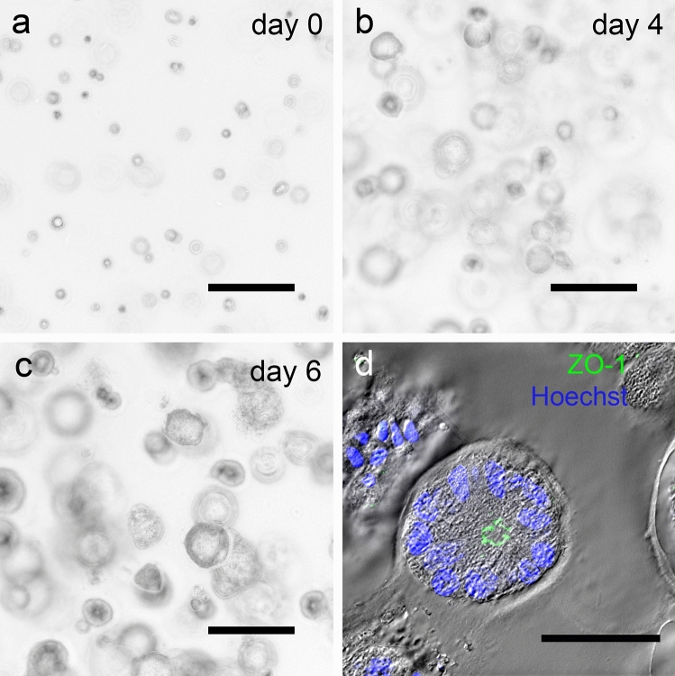
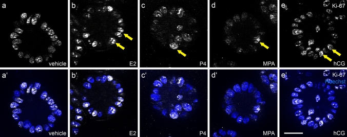
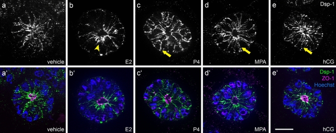
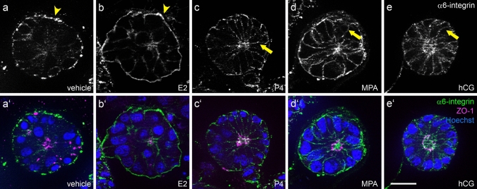
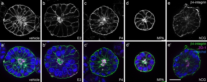
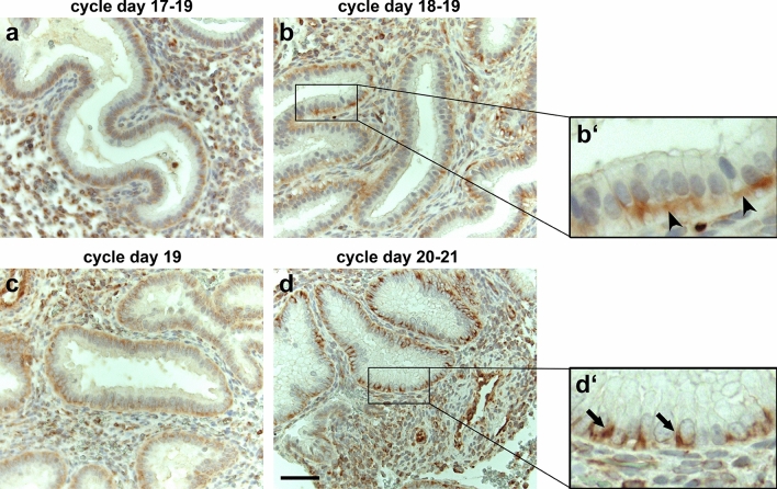
In human glandular endometrial epithelial cells, desmosomal and adherens junction proteins have been shown to extend from a subapically restricted lateral position to the entire lateral membrane during the implantation window of the menstrual cycle. Similarly, a menstrual cycle stage-dependent redistribution of the extracellular matrix adhesion protein α6-integrin has been reported. These changes are believed to be important for endometrial receptiveness and successful embryo implantation. To prove the hypothesis that steroid hormones and human choriogonadotropin can induce the redistribution of these adhesion molecules, we used the human endometrial cell line Ishikawa in a 3D culture system. Gland-like spheroids were grown in reconstituted basement membrane (Matrigel™). The lumen-bearing spheroids were treated for 2 or 4 days with ovarian steroids or human choriogonadotropin and then assessed by immunofluorescence microscopy. In addition, human endometrial biopsies were obtained from patients, who were in therapy for assisted reproductive technology, and were examined in parallel. Lateral redistribution of the desmosomal plaque protein desmoplakin 1 was observed in the spheroids treated either with progesterone, medroxyprogesterone acetate or human choriogonadotropin. Furthermore, the extracellular matrix adhesion protein α6-integrin showed an increased lateral membrane localization upon gestagen stimulation in the 3D culture system. The results of this study demonstrate that the 3D endometrial Ishikawa cell culture might be suited as an experimental model system to prove the effect of hormonal changes like those occurring during the window of implantation.
Keywords: 3D cell culture system; Cell adhesion; Endometrial receptivity; Epithelial polarity; Human endometrium; Ishikawa cell line.
Conflict of interest statement
The authors have no conflicts of interest to declare that are relevant to the content of this article.
Figures
Similar articles
-
Interaction of human trophoblast cells with gland-like endometrial spheroids: a model system for trophoblast invasion.Hum Reprod. 2015 Apr;30(4):906-16. doi: 10.1093/humrep/dev011. Epub 2015 Feb 5. Hum Reprod. 2015. PMID: 25662813
-
Redistribution of adhering junctions in human endometrial epithelial cells during the implantation window of the menstrual cycle.Histochem Cell Biol. 2012 Jun;137(6):777-90. doi: 10.1007/s00418-012-0929-0. Epub 2012 Feb 12. Histochem Cell Biol. 2012. PMID: 22327832
-
Studies using an in vitro model show evidence of involvement of epithelial-mesenchymal transition of human endometrial epithelial cells in human embryo implantation.J Biol Chem. 2012 Feb 10;287(7):4441-50. doi: 10.1074/jbc.M111.286138. Epub 2011 Dec 15. J Biol Chem. 2012. PMID: 22174415 Free PMC article.
-
Impact of ovarian hyperstimulation on the luteal phase.J Reprod Fertil Suppl. 2000;55:101-8. J Reprod Fertil Suppl. 2000. PMID: 10889839 Review.
-
Obesity and PCOS: the effect of metabolic derangements on endometrial receptivity at the time of implantation.Reprod Sci. 2015 Jan;22(1):6-14. doi: 10.1177/1933719114561552. Epub 2014 Dec 7. Reprod Sci. 2015. PMID: 25488942 Free PMC article. Review.
Cited by
-
The modeling of human implantation and early placentation: achievements and perspectives.Hum Reprod Update. 2025 Mar 1;31(2):133-163. doi: 10.1093/humupd/dmae033. Hum Reprod Update. 2025. PMID: 39673726 Free PMC article. Review.
-
An Assessment of the Mechanophysical and Hormonal Impact on Human Endometrial Epithelium Mechanics and Receptivity.Int J Mol Sci. 2024 Mar 27;25(7):3726. doi: 10.3390/ijms25073726. Int J Mol Sci. 2024. PMID: 38612536 Free PMC article.
-
Differential expression of tsRNAs and miRNAs in embryo culture medium: potential impact on embryo implantation.J Assist Reprod Genet. 2024 Mar;41(3):781-793. doi: 10.1007/s10815-024-03034-8. Epub 2024 Jan 25. J Assist Reprod Genet. 2024. PMID: 38270749 Free PMC article.
-
Changes in Epithelial Cell Polarity and Adhesion Guide Human Endometrial Receptivity: How In Vitro Systems Help to Untangle Mechanistic Details.Biomolecules. 2025 Jul 22;15(8):1057. doi: 10.3390/biom15081057. Biomolecules. 2025. PMID: 40867502 Free PMC article. Review.
-
In focus in HCB.Histochem Cell Biol. 2021 May;155(5):525-528. doi: 10.1007/s00418-021-01991-0. Epub 2021 May 11. Histochem Cell Biol. 2021. PMID: 33977373 No abstract available.
References
-
- Bielfeld AP, Pour SJ, Poschmann G, Stuhler K, Krussel JS, Baston-Bust DM. A proteome approach reveals differences between fertile women and patients with repeated implantation failure on endometrial level(-)does hCG render the endometrium of RIF patients? Int J Mol Sci. 2019 doi: 10.3390/ijms20020425. - DOI - PMC - PubMed
MeSH terms
Substances
Grants and funding
LinkOut - more resources
Full Text Sources
Other Literature Sources
