

Simultaneous Examination of Eosinophil Infiltration in Esophageal Mucosa and Muscle in Patients with Achalasia: Direct Biopsy of the Esophageal Muscle at Per-oral Endoscopic Myotomy
- PMID: 33502676
- PMCID: PMC7838844
- DOI: 10.1007/s10620-021-06827-4
Simultaneous Examination of Eosinophil Infiltration in Esophageal Mucosa and Muscle in Patients with Achalasia: Direct Biopsy of the Esophageal Muscle at Per-oral Endoscopic Myotomy
Abstract
Background: The relationship between eosinophilic esophagitis (EoE) and achalasia is not completely understood. There have been reports of eosinophilic infiltration of all esophageal layers in patients with achalasia. However, a routine endoscopic biopsy of the muscular layer is usually not feasible. We evaluate the safety and efficacy of muscle layer biopsy during per-oral endoscopic myotomy (POEM) as well as the prevalence of eosinophilic infiltration of the esophageal mucosa and muscular layer in patients with achalasia.
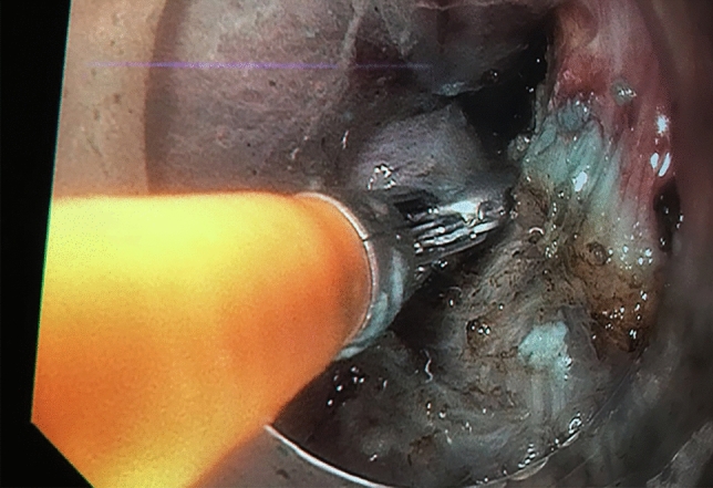
Patients and methods: All enrolled patients had diagnosed achalasia and had simultaneous biopsies of the muscular layer at the middle esophagus and distal esophageal sphincter as well as the mucosal layer of the proximal and distal esophagus during POEM. All POEM procedures took place from August 2018 to December 2018 or September 2019 to November 2019. Various demographic, disease-related, and procedure-related data were collected from chart review. Eosinophilic infiltration in the biopsy specimen was examined.
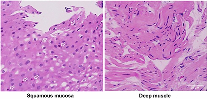
Key results: Twenty consecutive patients (65% female, age range: 21-84) with a pre-procedure Eckardt score of >6 were enrolled during the study period, with the duration of their achalasia ranging from 1 to 32 years. Eighteen patients had clinical symptomatic improvement after POEM, as defined by an Eckardt score <3. Endoscopic examination did not reveal any signs of eosinophilic esophagitis. Pathologic examination of biopsies revealed eosinophilic infiltration in three of 20 patients (15%) in the distal esophageal mucosa (all <15 eosinophils/HPF) and none in the proximal esophageal mucosa. There was no eosinophilic infiltration in the distal esophageal sphincter and the middle esophageal muscle. No complication was noted due to muscle biopsy.
Conclusions and inferences: Submucosal tunneling during POEM provides a safe access for direct esophageal muscle biopsy. This is the first report of the simultaneous biopsy of the esophageal mucosa and muscle in patients with achalasia. Contrary to all previously published studies, the association of esophageal eosinophilic infiltration and achalasia was not observed in this small sample study. Based on our findings, immune or autoimmune reaction rather than direct eosinophilic infiltration in the muscle is more likely the cause of achalasia.
Keywords: Achalasia; Eosinophilic esophagitis (EoE); POEM; Pathophysiology.
© 2021. The Author(s), under exclusive licence to Springer Science+Business Media, LLC part of Springer Nature.
Conflict of interest statement
Qiang Cai, MD, PhD, FACG, is a guarantor of the article. Specific author contributions: Huimin Chen, MD, PhD, searched the literature and wrote the original draft; Lucie F. Calderon, MD, Rushikesh Shah, MD, Wei Zheng, MD, Yue Xue, MD, and Steve Keilin, MD, searched the endoscopic photographs, revised the manuscript, and approved the final draft. Liang Xia, MD, PhD, Wei Wang, MD, Lianyong Li, MD, PhD, Baiwen Li, MD, PhD, and Steve Keilin, MD, revised the original draft and approved the final draft. Qiang Cai, MD, PhD, FACG, designed the concept and format of the article, provided endoscopic photographs, and revised and approved the final draft.
Figures
References
-
- Pandolfino JE, Kahrilas PJ, American Gastroenterological A. American Gastroenterological Association medical position statement: clinical use of esophageal manometry. Gastroenterology 2005;128: 207–8. - PubMed
MeSH terms
LinkOut - more resources
Full Text Sources
Other Literature Sources
Medical
