

Ginsenoside Rb1 Facilitates Browning by Repressing Wnt/β-Catenin Signaling in 3T3-L1 Adipocytes
- PMID: 33503016
- PMCID: PMC7849207
- DOI: 10.12659/MSM.928619
Ginsenoside Rb1 Facilitates Browning by Repressing Wnt/β-Catenin Signaling in 3T3-L1 Adipocytes
Abstract
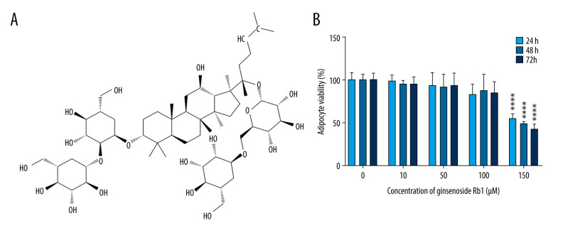
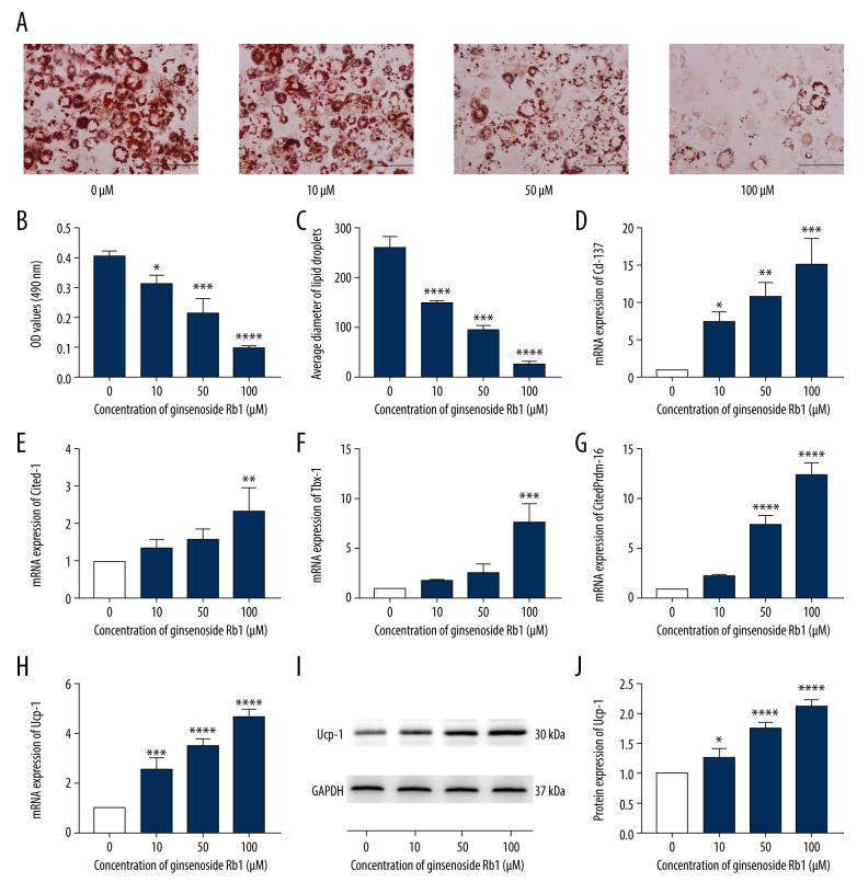
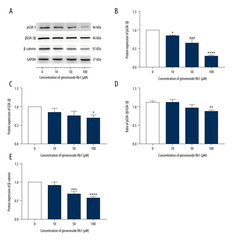
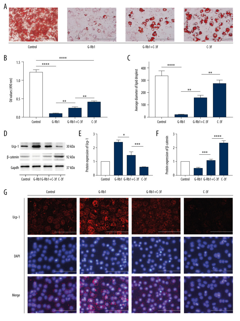
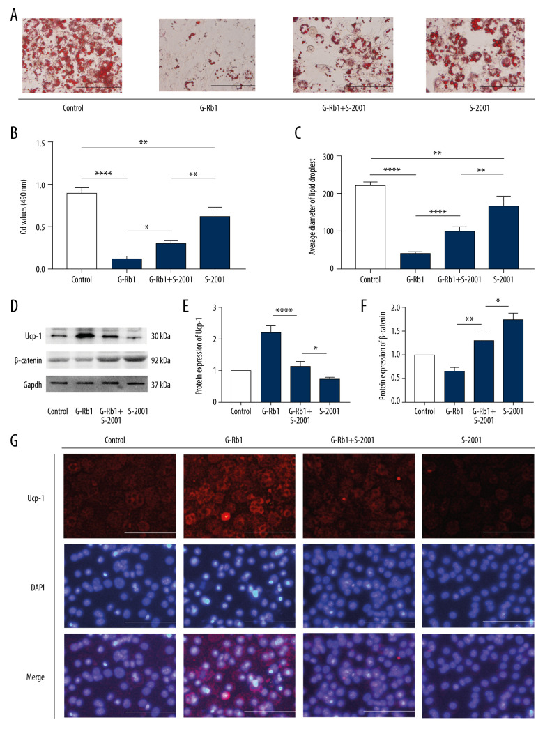
BACKGROUND The discovery of browning in white adipose tissue has provided new ideas for treating obesity. Many studies have reported that ginsenoside Rb1 (G-Rb1) has activity against diabetes, inflammation, and obesity, but further investigation is needed on the effect and mechanism of G-Rb1 on browning. MATERIAL AND METHODS We treated 3T3-L1 adipocytes with 0-200 μM G-Rb1, and 0.5 μM Compound 3f and 30 μM SKL2001 were used to activate Wnt/b-catenin signaling. Adipocyte activity was evaluated by Cell Counting Kit-8. Oil Red O staining was used to detect the lipid droplets. Quantitative real-time polymerase chain reaction was used to measure the expression of Cd-137, Cited-1, Txb-1, Prdm-16, and Ucp-1 mRNA. Western blotting was used to measure the expression of Ucp-1, pGSK-3ß (Ser 9), GSK- 3ß, and ß-catenin proteins. The expression of Ucp-1 was also detected with immunofluorescence. RESULTS Adipocyte activity was not affected by 0-100 μM G-Rb1. However, G-Rb1 dose-dependently reduced the accumulation of lipid droplets; increased the expression of Cd-137, Cited-1, Txb-1, Prdm-16, and Ucp-1 mRNA; and increased the expression of Ucp-1, pGSK-3ß (Ser 9), GSK-3ß, and ß-catenin proteins. The accumulation of lipid droplets and the expression of Ucp-1 protein decreased as b-catenin increased. CONCLUSIONS G-Rb1 at various concentrations (0-100 μM) promoted the browning of adipocytes in a dose-dependent manner. Further, we confirmed that activation of Wnt/ß-catenin signaling could inhibit browning. Therefore, the browning promoted by G-Rb1 may be associated with the inhibition of Wnt/ß-catenin signaling.
Conflict of interest statement
None.
Figures
Similar articles
-
Ginsenoside Rb1 promotes browning through regulation of PPARγ in 3T3-L1 adipocytes.Biochem Biophys Res Commun. 2015 Oct 23;466(3):530-5. doi: 10.1016/j.bbrc.2015.09.064. Epub 2015 Sep 14. Biochem Biophys Res Commun. 2015. PMID: 26381176
-
Black Ginseng and Ginsenoside Rb1 Promote Browning by Inducing UCP1 Expression in 3T3-L1 and Primary White Adipocytes.Nutrients. 2019 Nov 12;11(11):2747. doi: 10.3390/nu11112747. Nutrients. 2019. PMID: 31726767 Free PMC article.
-
Ginsenoside Rg3 Induces Browning of 3T3-L1 Adipocytes by Activating AMPK Signaling.Nutrients. 2020 Feb 7;12(2):427. doi: 10.3390/nu12020427. Nutrients. 2020. PMID: 32046061 Free PMC article.
-
Sijunzi decoction improves lipid metabolism via regulation of Wnt/β-catenin signaling pathway in diabetic mice and 3T3-L1 cells.J Ethnopharmacol. 2025 Apr 25;346:119672. doi: 10.1016/j.jep.2025.119672. Epub 2025 Mar 24. J Ethnopharmacol. 2025. PMID: 40139576
-
Critical review on anti-obesity effects of phytochemicals through Wnt/β-catenin signaling pathway.Pharmacol Res. 2022 Oct;184:106461. doi: 10.1016/j.phrs.2022.106461. Epub 2022 Sep 21. Pharmacol Res. 2022. PMID: 36152739 Review.
Cited by
-
Natural products for treatment of premature ovarian failure: a narrative review.J Tradit Chin Med. 2023 Jun;43(3):606-617. doi: 10.19852/j.cnki.jtcm.20230227.002. J Tradit Chin Med. 2023. PMID: 37147765 Free PMC article. Review.
-
Ginseng-derived compounds as potential anticancer agents targeting cancer stem cells.J Ginseng Res. 2024 May;48(3):266-275. doi: 10.1016/j.jgr.2024.03.003. Epub 2024 Mar 12. J Ginseng Res. 2024. PMID: 38707642 Free PMC article. Review.
-
Signaling pathways in obesity: mechanisms and therapeutic interventions.Signal Transduct Target Ther. 2022 Aug 28;7(1):298. doi: 10.1038/s41392-022-01149-x. Signal Transduct Target Ther. 2022. PMID: 36031641 Free PMC article. Review.
-
Lycopene and Garcinia cambogia Induce White-to-Brown Adipose Differentiation: An Innovative Strategy to Curb Obesity.Pharmaceuticals (Basel). 2024 Jul 25;17(8):986. doi: 10.3390/ph17080986. Pharmaceuticals (Basel). 2024. PMID: 39204091 Free PMC article.
-
Targeting Adenosine Receptor by Polydeoxyribonucleotide: An Effective Therapeutic Strategy to Induce White-to-Brown Adipose Differentiation and to Curb Obesity.Pharmaceuticals (Basel). 2021 Jul 27;14(8):728. doi: 10.3390/ph14080728. Pharmaceuticals (Basel). 2021. PMID: 34451825 Free PMC article.
References
-
- Cuthbert K, Hardin S, Zelkowitz R, Mitchell K. Eating disorders and overweight/obesity in veterans: Prevalence, risk factors, and treatment considerations. Curr Obes Rep. 2020;9(2):98–108. - PubMed
-
- Yang G, Zhuo J, Lin Y, et al. Ginsenoside Rb1 prevents dysfunction of endothelial cells by suppressing inflammatory response and apoptosis in the high-fat diet plus balloon catheter-injured rabbit model via the G protein-coupled estrogen receptor-mediated phosphatidylinositol 3-kinases (PI3K)/Akt pathway. Med Sci Monit. 2019;25:7407–17. - PMC - PubMed
MeSH terms
Substances
LinkOut - more resources
Full Text Sources
Other Literature Sources
Research Materials
Miscellaneous
