. 2021 Jan 20;35(12):e351204.
doi: 10.1590/ACB351204. eCollection 2021.

Evaluation of the effects of diode laser application on experimental orthodontic tooth movements in rats. Histopathological analysis

Affiliations

Evaluation of the effects of diode laser application on experimental orthodontic tooth movements in rats. Histopathological analysis

Mehmet Ali Karabel et al. Acta Cir Bras. .

Abstract

Purpose: To evaluate the effect of diode laser use on experimental orthodontic tooth movements.

Methods: Thirty Rattus norvegicus albinus Wistar were divided into three equal groups (n = 10), two experimentals and one control. Applying 20 g orthodontic force were attached to the maxillary incisors of the rats in all groups. Low dose laser was applied to the surrounding tissues of the maxillary incisors of the rats in the experimental groups. Two exposure times for laser irradiation were used for seven days: t = 12 min (energy dose = 72 J) and t = 9 min (energy dose = 54 J) by a 0.1 W DEKA brand diode laser with wavelength of 980 nm.

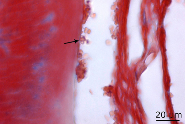
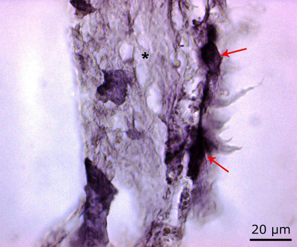
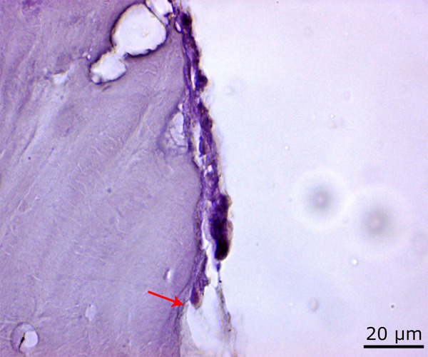
Results: Osteoclastic activation increased in the 72 J group when compared to control group and decreased in comparison to the 54 J group. Osteoblastic activation was decreased in the 72 J group when compared to the control group and increased in comparison to the 54 J group.

Conclusions: Applying 54 J laser energy has been found effective to accelerate the orthodontic tooth movement.


Conflict of interest statement

Conflict of interest:none

Figures

Figure 1
Figure 1. Passive configuration of appliance.
Figure 2
Figure 2. Active configuration of appliance.
Figure 3
Figure 3. Intraoral view of the appliance applied to the experimental animal.
Figure 4
Figure 4. Normal osteoblast activity (red arrow) in the control group (H&E, ×100).
Figure 5
Figure 5. Normal osteoclast activity (black arrow) in the control group (H&E, ×100).
Figure 6
Figure 6. Increased osteoclast activity in the 54 J laser group (black arrow) and regions where bone destruction is intense (*) (H&E, ×100).
Figure 7
Figure 7. Decrease in osteoblast activity in the 54 J laser group (black arrow) (Masson’s trichrome staining, ×100).
Figure 8
Figure 8. Increase in osteoclast activity in the 54 J laser group (red arrow) and density in the areas of bone degeneration (*) (PAS, ×100).
Figure 9
Figure 9. Osteoclast activity (red arrow) in the 72 J laser group. Osteoblastic activity (black arrow), which showed an increase when compared to the 54 J group and decrease when compared to the control group. Also, partial normalization in bone degeneration (*) (H&E, ×100).
Figure 10
Figure 10. Increased osteoblastic activity in the 72 J laser group when compared to the 54 J group (black arrow) (Masson’s trichrome staining ×100).
Figure 11
Figure 11. Increased osteoclastic activity in the 72 J laser group compared to the control group (red arrow) (PAS, ×100).

Similar articles

Cited by

References

    1. Goulart CS, Nouer PR, Mouramartins L, Garbin IU, de Fátima. Photoradiation and orthodontic movement: experimental study with canines. Photomed Laser Surg. 2006;24(2):192–196. doi: 10.1089/pho.2006.24.192. - DOI - PubMed
    1. Khouw FE, Goldhaber P. Changes in vasculature of periodontium associated with tooth movement in the rhesus monkey and dog. Arch Oral Biol. 1970;15(12):1125–1132. doi: 10.1016/0003-9969(70)90003-8. - DOI - PubMed
    1. Furstman L, Bernick S, Aldrich D. Differential response incident to tooth movement. Am J Orthod. 1971;59(6):600–608. doi: 10.1016/0002-9416(71)90007-8. - DOI - PubMed
    1. Rygh P, Bowling K, Hovlandsdal L, Williams S. Activation of the vascular system: a main mediator of periodontal fiber remodeling in orthodontic tooth movement. Am J Orthod. 1986;89(6):453–468. doi: 10.1016/0002-9416(86)90001-1. - DOI - PubMed
    1. Engström C, Granström G, Thilander B. Effect of orthodontic force on periodontal tissue metabolism. A histologic and biochemical study in normal and hypocalcemic young rats. Am J Orthod Dentofacial Orthop. 1988;93(6):486–495. doi: 10.1016/0889-5406(88)90077-7. - DOI - PubMed