. 2021 Feb 22;49(3):1737-1748.
doi: 10.1093/nar/gkaa1276.

First insights into the structural features of Ebola virus methyltransferase activities

Affiliations

First insights into the structural features of Ebola virus methyltransferase activities

Coralie Valle et al. Nucleic Acids Res. .

Abstract

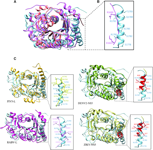
The Ebola virus is a deadly human pathogen responsible for several outbreaks in Africa. Its genome encodes the 'large' L protein, an essential enzyme that has polymerase, capping and methyltransferase activities. The methyltransferase activity leads to RNA co-transcriptional modifications at the N7 position of the cap structure and at the 2'-O position of the first transcribed nucleotide. Unlike other Mononegavirales viruses, the Ebola virus methyltransferase also catalyses 2'-O-methylation of adenosines located within the RNA sequences. Herein, we report the crystal structure at 1.8 Å resolution of the Ebola virus methyltransferase domain bound to a fragment of a camelid single-chain antibody. We identified structural determinants and key amino acids specifically involved in the internal adenosine-2'-O-methylation from cap-related methylations. These results provide the first high resolution structure of an ebolavirus L protein domain, and the framework to investigate the effects of epitranscriptomic modifications and to design possible antiviral drugs against the Filoviridae family.


Figures

Figure 1.
Figure 1.
SUDV MTase structure resolved by X-ray crystallography. (A) Crystal structure of the Sudan ebolavirus (SUDV) methyltransferase (MTase) domain (blue) in complex with a VHH (orange). The N- and C-terminal extremities are indicated by the N and C letters, respectively, in both proteins. The catalytic residues K1813, D1924, K1959 and E1996 of the SUDV MTase are highlighted in red. The SUDV MTase structure shows the presence of eight β-sheets alternated with eight α-helices and two undefined loops (residues K1764 to P1775 and residues V1795 to S1808, dashed lines). The VHH adopts a classic β-sandwich fold structure composed of eight strands connected by flexible loops. Two loops (residues G55 to A61 and residues A100 to Y109) and a turn (residues R27 to R31) are involved in the interaction with the SUDV MTase domain, at the opposite side of the catalytic pocket. (B) Topological organization of the SUDV MTase domain (bottom) and of the canonical Rossmann fold of a SAM-dependent MTase (top). The MTase Rossmann fold is defined by a β-α-β motif (here β2-α3-β3) that contacts the SAM methyl donor. The overall strand/helix architecture is boxed in gray in the SUDV MTase fold representation. Helices are depicted as cyan barrels, β-strands as blue arrows, and coils as black lines. The N- and C-terminal extremities are indicated by N and C, respectively. Red points correspond to catalytic residues within the Rossmann fold secondary structure. This representation highlights the additional features at the N-terminus (1 β-strand and 1 α-helix) and C-terminus (1 α-helix) of the SUDV MTase.
Figure 2.
Figure 2.
Identification of conserved functional pockets. (A) Modeling of the S-adenosylmethionine (SAM) molecule (red) within the SUDV MTase structure based on the hMPV MTase structure co-crystallized with SAM. Residues involved in the SAM-binding pocket (SAMP) are in orange, and catalytic residues in yellow. (B) Superimposition of the SUDV MTase domain (gray) with the hMPV L MTase domain (PDB: 4UCI, salmon), with the SAM molecule in red. The flexible loop participating in SAMP is highlighted in orange in the hMPV and SUDV MTase structures. The hMPV loop adopts a ‘closed’ conformation, clamping the SAM substrate in SAMP, whereas the SUDV loop shows an ‘open’ conformation. (C) Modeling of a GTP molecule (red) within the SUDV MTase structure based on the hMPV MTase structure co-crystallized with GTP. Residues involved in the substrate-binding pocket (SUBP) as well as the deep hydrophobic cavity (NSP) are in pink and catalytic residues are in yellow.
Figure 3.
Figure 3.
Single-mutation analysis of the SAM-binding and RNA-binding pockets. (A) MTase activity of SUDV WT and mutated MTase+CTD in the SAMP. The G1835S, G1837S, T1854A and L1855A mutations led to complete loss of the tested activities (cap-N7, cap-2′-O and internal A-2′-O-methylations). Mutation E1834A promoted the three methylation activities. (B) MTase activity of SUDV WT and mutated MTase+CTD in the SUBP and NSP. The Y1800A and S1809A mutations in the putative RNA-binding groove led to loss overall MTase activities (cap-N7, cap-2′-O and internal A-2′-O-methylations). Other mutations, such as I1806A, V1807A, T1927A and S1990A possibly involved in RNA binding, showed a significant reduction of all MTase activities. Several mutations in SUBP resulted in the uncoupling of the different MTase activities. The S1991A and K1993A mutations led to a drastic reduction (approximately by 50%) and almost complete loss of 2′-O-MTase activities (cap and internal A-2′-O-MTase activities), respectively, but not of the N7 MTase activity. The S1808A and R1792A mutations similarly impaired only the internal A-2′-O-MTase activity, but not the MTase activities associated with cap synthesis. Data are the mean ± standard deviation (n = 3); *P< 0.05, **P< 0.01, ***P< 0.001 (two-way ANOVA and multiple comparison Dunnett test, WT versus mutation).
Figure 4.
Figure 4.
Structural comparison with other viral MTase domains. (A) Superimposition of the Sudan ebolavirus (SUDV) methyltransferase domain (MTase, sky blue) with the human metapneumovirus (hMPV) L MTase domain (PDB: 4UCI, salmon) and vesicular stomatitis virus (VSV) L protein (PDB: 5A22, purple). Compared with the other mononegaviruses, the additional N-terminal α-helix in SUDV MTase (α1) is not found in the hMPV and VSV MTase structures. (B) Close-up of the α1 homologous region in VSV superimposed to SUDV α1 in which the positively charged and polar residues are identified. This region was not resolved in the hMPV MTase structure, suggesting high flexibility. (C) Structural comparison with other viral MTase domains. Superimposition of SUDV MTase (sky blue) with the parainfluenza 5 virus L protein (PIV5-L, PDB: 6V85, top left, yellow), rabies virus L protein (RABV-L, PDB: 6UEB, bottom left, dark pink), dengue 2 virus non-structural protein 5 (DENV2-NS5, PDB: 5ZQK, top right, dark khaki) and Zika virus NS5 (ZIKV-NS5, PDB: 5M5B, bottom right, light khaki) MTase domains. The supplementary N-terminal α-helix in SUDV MTase (α1) was found also in the DENV2 and ZIKV NS5 MTase structures (red). A close-up of the α1 homologous regions in all structures is represented on the left of each superimposition with the positively charged and polar residues identified.
Figure 5.
Figure 5.
Structural mapping of residues involved in SUDV cap and internal methylations. Mapping of single mutations in the SUDV MTase domain (grey), coloured according to their involvement in cap-N7 methylation (left, residues in green), cap-2′-O-methylation (middle, residues in dark blue), and internal A-2′-O-methylation (right, in dark blue). The catalytic site (K1813-D1924-K1959-E1996) is coloured in pink and a SAM molecule (yellow) was modelled according to its putative position for the different methyltransferase activities.

Similar articles

Cited by

References

    1. Lawrence P., Danet N., Reynard O., Volchkova V., Volchkov V.. Human transmission of Ebola virus. Curr. Opin. Virol. 2017; 22:51–58. - PubMed
    1. Ollmann Saphire E. A Vaccine against Ebola Virus. Cell. 2020; 181:6. - PubMed
    1. Elliott L.H., Sanchez A., Holloway B.P., Kiley M.P., McCormick J.B.. Ebola protein analyses for the determination of genetic organization. Arch. Virol. 1993; 133:423–436. - PubMed
    1. Sanchez A., Kiley M.P., Holloway B.P., Auperin D.D.. Sequence analysis of the Ebola virus genome: organization, genetic elements, and comparison with the genome of Marburg virus. Virus Res. 1993; 29:215–240. - PubMed
    1. Ogino T., Banerjee A.K.. Unconventional mechanism of mRNA capping by the RNA-dependent RNA polymerase of vesicular stomatitis virus. Mol. Cell. 2007; 25:85–97. - PubMed

Publication types