. 2021 May 7;49(8):e45.
doi: 10.1093/nar/gkab011.

A non-radioactive, improved PAR-CLIP and small RNA cDNA library preparation protocol

Affiliations

A non-radioactive, improved PAR-CLIP and small RNA cDNA library preparation protocol

Dimitrios G Anastasakis et al. Nucleic Acids Res. .

Abstract

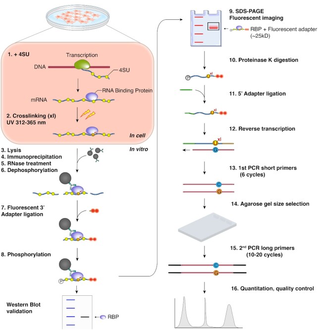
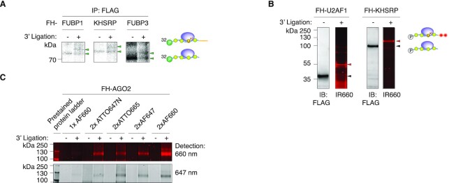
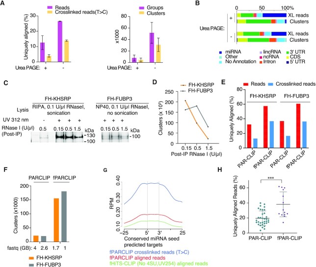
Crosslinking and immunoprecipitation (CLIP) methods are powerful techniques to interrogate direct protein-RNA interactions and dissect posttranscriptional gene regulatory networks. One widely used CLIP variant is photoactivatable ribonucleoside enhanced CLIP (PAR-CLIP) that involves in vivo labeling of nascent RNAs with the photoreactive nucleosides 4-thiouridine (4SU) or 6-thioguanosine (6SG), which can efficiently crosslink to interacting proteins using UVA and UVB light. Crosslinking of 4SU or 6SG to interacting amino acids changes their base-pairing properties and results in characteristic mutations in cDNA libraries prepared for high-throughput sequencing, which can be computationally exploited to remove abundant background from non-crosslinked sequences and help pinpoint RNA binding protein binding sites at nucleotide resolution on a transcriptome-wide scale. Here we present a streamlined protocol for fluorescence-based PAR-CLIP (fPAR-CLIP) that eliminates the need to use radioactivity. It is based on direct ligation of a fluorescently labeled adapter to the 3'end of crosslinked RNA on immobilized ribonucleoproteins, followed by isolation of the adapter-ligated RNA and efficient conversion into cDNA without the previously needed size fractionation on denaturing polyacrylamide gels. These improvements cut the experimentation by half to 2 days and increases sensitivity by 10-100-fold.


Figures

Figure 1.
Figure 1.
fPAR-CLIP schematic.
Figure 2.
Figure 2.
Ligation of a 3′ adapter to crosslinked RNPs results in a predictable electrophoretic mobility shift. (A) SDS-PAGE of crosslinked, radiolabled, and immunoprecipitated FLAG-HA-tagged (FH) FUBP1, FH-KHSRP, and FH-FUBP3 RNPs before and after ligation to a 3′ adapter. (B) Immunoblots and fluorescent images of SDS-PAGE separating fluorescent adapter-ligated crosslinked FH-U2AF1 (left panels) and FH-KHSRP (right panels) RNPs. Fluorescent images were detected at 700 nm wavelength. (C) Comparison of FH-AGO2 fPAR-CLIP band intensity using different fluorescent adapters labeled with the indicated dyes and imaged at 647 or 660 nm detection wavelength.
Figure 3.
Figure 3.
Streamlined cDNA library construction in fPAR-CLIP increases cDNA library complexity. (A) Left panel: Percentage of uniquely aligned reads from PAR-CLIP libraries size selected after 5′adapter ligation by denaturing polyacrylamide gel electrophoresis (DPAGE) or used directly as input in RT and low cycle PCR followed by size selection by automated agarose gel electrophoresis. Right panel: comparison of groups and clusters identified in both libraries. (B) Distribution of crosslink reads across different annotation categories for the samples from (A). (C) Fluorescent image of SDS PAGE using different concentration of RNase I after KHSRP and FUBP3 IP. (D) Number of clusters for FH-KHSRP and FH-FUBP3 fPAR-CLIPs from (C) using different concentration of RNase I. (E) Percentage of uniquely aligned sequence reads (red bars) or crosslinked reads (blue bars) from PAR-CLIP and fPAR-CLIP experiments for FH-KHSRP and FUBP3. (F) Number of binding sites identified from PAR-CLIP and fPAR-CLIP experiments for FH-KHSRP (orange) and FH-FUBP3 (grey). The size of the raw sequencing file is indicated below. (G) Enrichment of FH-AGO2 fPARCLIP and HiTS-CLIP footprints across predicted, conserved miRNA target sites. (H) Comparison of uniquely aligned reads from all published PAR-CLIP libraries with at least 20,000 RBP binding sites compared to fPAR-CLIP libraries produced in our laboratory (* P < 0.05, Mann–Whitney U test). See Supplementary Table S2 for the list of analyzed PAR-CLIP and fPAR-CLIP experiments.

References

    1. Singh G., Pratt G., Yeo G.W., Moore M.J.. The clothes make the mRNA: past and present trends in mRNP fashion. Annu. Rev. Biochem. 2015; 84:325–354. - PMC - PubMed
    1. Gerstberger S., Hafner M., Tuschl T.. A census of human RNA-binding proteins. Nat. Rev. Genet. 2014; 15:829–845. - PMC - PubMed
    1. Gerstberger S., Hafner M., Ascano M., Tuschl T.. Evolutionary conservation and expression of human RNA-binding proteins and their role in human genetic disease. Adv. Exp. Med. Biol. 2014; 825:1–55. - PMC - PubMed
    1. Ray D., Kazan H., Chan E.T., Pena Castillo L., Chaudhry S., Talukder S., Blencowe B.J., Morris Q., Hughes T.R.. Rapid and systematic analysis of the RNA recognition specificities of RNA-binding proteins. Nat. Biotechnol. 2009; 27:667–670. - PubMed
    1. Lambert N., Robertson A., Jangi M., McGeary S., Sharp P.A., Burge C.B.. RNA Bind-n-Seq: quantitative assessment of the sequence and structural binding specificity of RNA binding proteins. Mol. Cell. 2014; 54:887–900. - PMC - PubMed

Publication types

MeSH terms