. 2021 Jan 26;34(4):108673.
doi: 10.1016/j.celrep.2020.108673.

Aspergillus fumigatus tryptophan metabolic route differently affects host immunity

Affiliations

Aspergillus fumigatus tryptophan metabolic route differently affects host immunity

Teresa Zelante et al. Cell Rep. .

Abstract

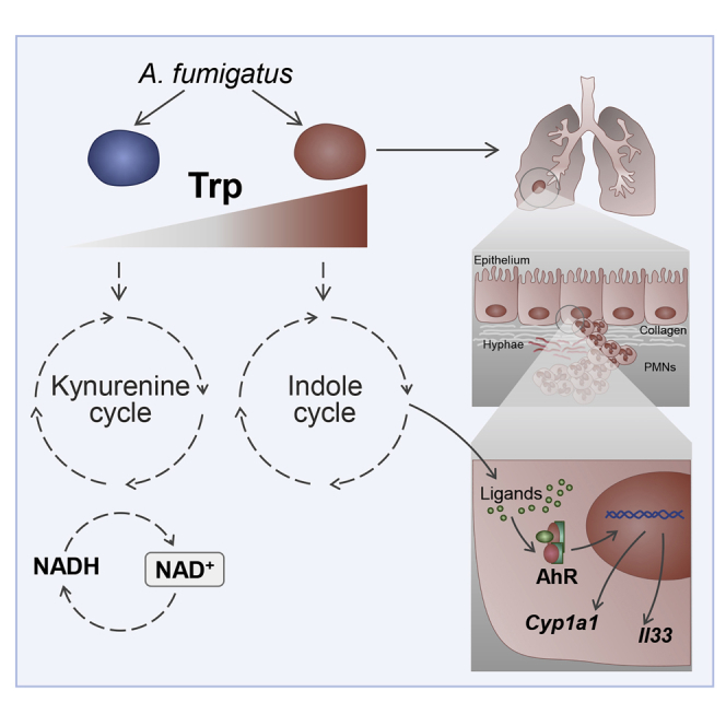
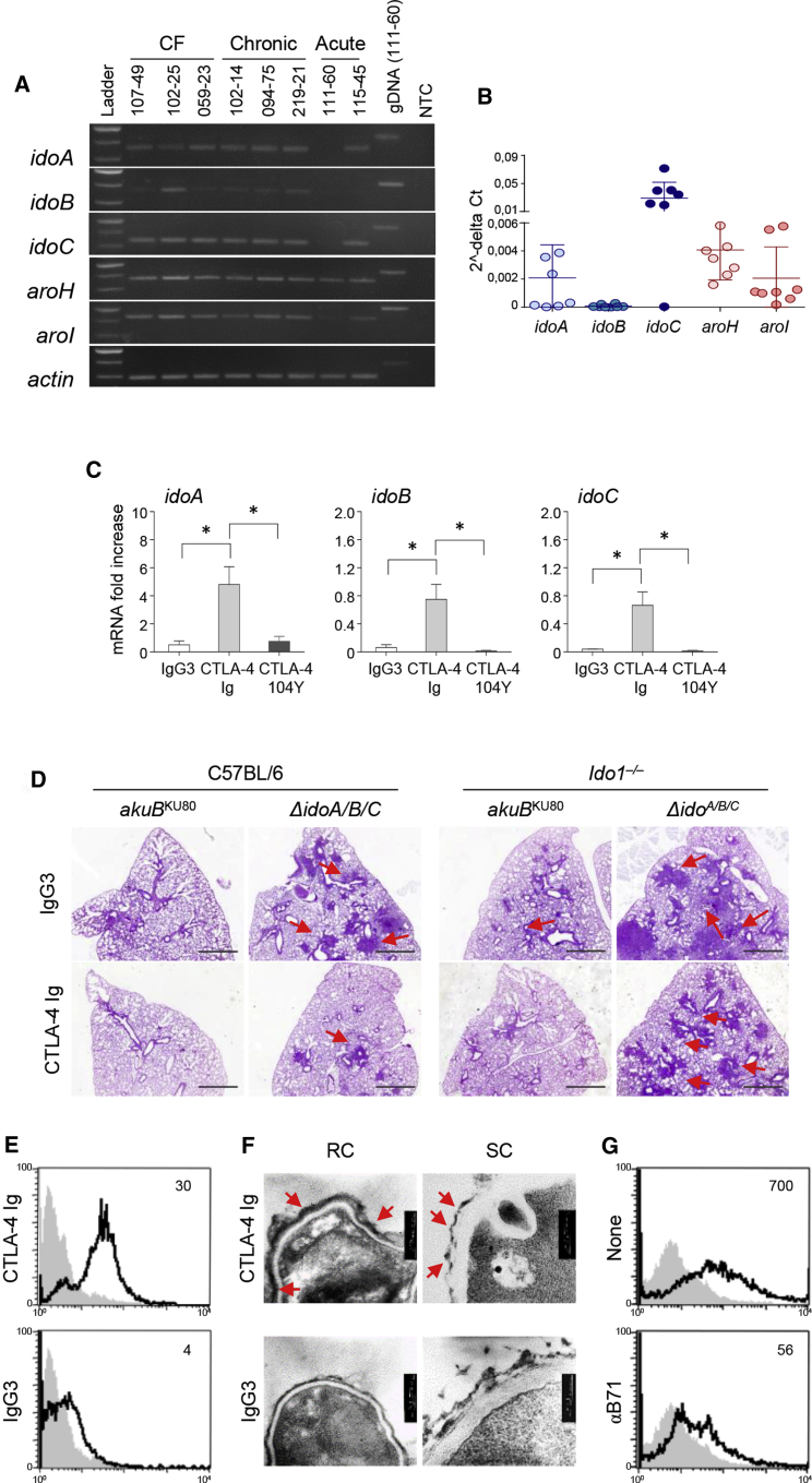
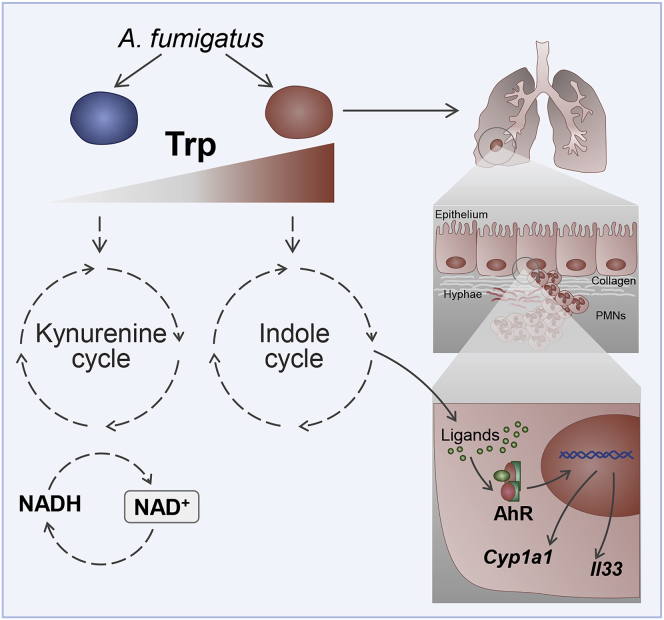
Indoleamine 2,3-dioxygenases (IDOs) degrade l-tryptophan to kynurenines and drive the de novo synthesis of nicotinamide adenine dinucleotide. Unsurprisingly, various invertebrates, vertebrates, and even fungi produce IDO. In mammals, IDO1 also serves as a homeostatic regulator, modulating immune response to infection via local tryptophan deprivation, active catabolite production, and non-enzymatic cell signaling. Whether fungal Idos have pleiotropic functions that impact on host-fungal physiology is unclear. Here, we show that Aspergillus fumigatus possesses three ido genes that are expressed under conditions of hypoxia or tryptophan abundance. Loss of these genes results in increased fungal pathogenicity and inflammation in a mouse model of aspergillosis, driven by an alternative tryptophan degradation pathway to indole derivatives and the host aryl hydrocarbon receptor. Fungal tryptophan metabolic pathways thus cooperate with the host xenobiotic response to shape host-microbe interactions in local tissue microenvironments.

Keywords: AhR; Aspergillus fumigatus; IDO; IL-33; NAD; aspergillosis; indoles; inflammation; tryptophan.


Conflict of interest statement

Declaration of interests The authors declare no competing interests.

Figures

None
Graphical abstract
Figure 1
Figure 1
Aspergillus Idos catabolize Trp for NAD+ and kynurenines biosynthesis (A) Maximum likelihood phylogenetic analysis of IDOs. (B) mRNA fold increase of Aspergillus idoA, idoB, and idoC in GMM and GMM + Trp at 24 h of culture. (C and D) Peak levels and relative abundance of l-kynurenine (Kyn) and nicotinamide (D) in WT (Aspergillus akuBKU80) and mutant strains inoculated on solid GMM supplemented with NAM and Trp and cultured at 37°C for 84 h in triplicates. (E) Growth of parental akuBKU80and mutant strains in GMM, GMM + Trp (60 μM), and NAD supplementation (NAM) to the growth medium. (B and D) Data are represented as mean ± SD. Graphs are representative of data collected from three independent replicate experiments. (B) Statistical significance (∗∗∗p < 0.0001) was determined against the untreated (GMM) (two-tailed Student’s t test unpaired parametric). (D) Statistical significance (∗∗∗p < 0.0001) was determined against the Aspergillus akuBKU80 strain (two-way ANOVA and Bonferroni’s akuBKU80 versus ΔidoA/B and versus ΔidoA/B/C). (C and E) Experiments were repeated three times. See also Figures S1–S4.
Figure 2
Figure 2
Aspergillus Trp alternative metabolism: the Aro pathway (A and B) Pie diagram and peak levels of Trp-derived metabolites performed at 24 h of culture in GMM + Trp by targeted metabolomics of Aspergillus WT strain. (C) Trp metabolic pathways, enzymes, and molecular structures of Trp-indole derivatives. (D and E) RT-PCR of Aspergillus genes involved in the catabolic pathway (D) and relative abundance of indole-derivatives in WT (Aspergillus akuBKU80) and mutant strains inoculated on solid GMM supplement with NAM and Trp and cultured at 37°C for 84 h (E). (F) Radial growth of Aspergillus akuBKU80 and A. fumigatus aro mutants. Strains were inoculated with 104 conidia onto the solidified GMM supplemented with 5 mM l-Trp. Colony diameters of each strain were measured after 1, 3, and 5 days of growth at 37°C. (A, B, and D) Experiments were repeated three times. Statistical significance (∗∗p < 0.001) was determined against the Aspergillus akuBKU80 strain (unpaired t test: akuBKU80 versus ΔidoA/B and versus ΔidoA/B/C). See also Figures S4 and S5.
Figure 3
Figure 3
Ido deficiency increases A. fumigatus pathogenicity in vivo (A) Peak levels of Trp-derived metabolites performed at 24 h of culture in RPMI 1640 in normoxia or hypoxia by targeted metabolomics of Aspergillus WT strain. (B) mRNA expression (fold increase) of idoA, idoB, and idoC in Aspergillus grown in normoxia or hypoxia after 24 h of culture. (C–I) C57BL/6 mice (n = 9) were infected intranasally with 2 × 107 resting conidia of WT akuBKU80 or ΔidoA/B/C A. fumigatus mutant and sacrificed at different days postinfection (dpi). (C) mRNA fold change of idoA, idoB, and idoC at 7 and 14 dpi in the lung of C57BL/6 mice. (D) Survival rates of C57BL/6 mice. (E) 18S rRNA expression measured by qPCR from C57BL/6 lungs. (F) Bronchoalveolar lavage (BAL) cell count analysis of C57BL/6 showing the percentage of pulmonary neutrophils (PMNs). (G) Histopathological analyses (periodic acid-Schiff [PAS], Alcian blue, and Masson’s Trichrome) in C57BL/6 lung tissue at 7 dpi. Scale bars, 10 μm and 100 μm. (H) Total pathology score on lung histology at 7 dpi. (I) Cytokine levels by ELISA. (B, C, E, F, H, and I) Data are represented as means ± SD. Statistical significance for (B) (p < 0.01, ∗∗p < 0.001, ∗∗∗p < 0.0001) was determined against Aspergillus samples cultured in normoxia (Twotwo- tailed Student’s t Test test unpaired parametric). (C) Statistical significance, (∗∗p < 0.001) was determined against mice infected with the Aspergillus akuBKU80 strain at 7 dpi (Twotwo- tailed Student’s t Test test unpaired parametric). (D) Nonparametric test of infected mice with AkuBKU80 versus ΔidoA/B/C. (E and F) Statistical significance (p < 0.01, ∗∗p < 0.001) was determined against mice infected with the Aspergillus akuBKU80 strain (E) or naive mice (F) (two-way ANOVA and Bonferroni post hoc test). (H–I) Statistical significance (∗∗p < 0.001, ∗∗∗p < 0.0001) was determined against mice infected with the Aspergillus akuBKU80 strain (two-tailed Student’s t test unpaired parametric). For (I), statistical significance (∗∗p < 0.001) was determined against mice uninfected (naive) (Two two-tailed Student’s t Test test unpaired parametric). Micrographs are representative of three replicate experiments. See also Figures S6 and S7.
Figure 4
Figure 4
The AroH/I pathway promotes lung inflammation via AhR C57BL/6 and Ahr–/– mice (n = 9) were infected intranasally with 2 × 107 resting conidia of indicated A. fumigatus strains and sacrificed at 7 dpi. (A) Survival rates. (B) 18S rRNA expression measured by qPCR from C57BL/6 lungs. (C) Cytokine levels by ELISA. (D) mRNA fold change of aroH at 7 and 14 dpi in the lung of C57BL/6 mice. (E) mRNA fold change of Cyp1a1 at 7 dpi in the lung of C57BL/6 mice. (F) Survival rates of C57BL/6 and Ahr–/– mice. (G) 18S rRNA expression measured by qPCR from C57BL/6 lungs. (H) Cytokine levels by ELISA from C57BL/6 and Ahr–/– mouse lungs. (I) Histopathological analyses (PAS) in C57BL/6 lung tissue at 7 dpi. Scale bars, 10 μm. (J) Total pathology score on lung histology at 7 dpi. (K) 18S rRNA expression measured by qPCR from C57BL/6 lungs at 7 dpi. (L) Cytokine levels by ELISA at 7 dpi. Data are represented as means ± SD. Graphs are representative of data collected from three independent replicate experiments. (B, C, and E) Statistical significance (p < 0.01, ∗∗p < 0.001, ∗∗∗p < 0.0001) was determined against mice infected with the Aspergillus akuBKU80 strain (one-way ANOVA and Bonferroni post hoc test). (D, G, H, J–L) Statistical significance (∗∗p < 0.001, ∗∗∗p < 0.0001) was determined against mice infected with the Aspergillus akuBKU80 strain (two-way ANOVA and Bonferroni post hoc test). (E) statistical significance (∗∗∗p < 0.0001, ∗∗p < 0.001) was determined against mice infected with the Aspergillus akuBKU80 strain (one-way ANOVA and Bonferroni post hoc test). (F) Nonparametric test AkuBKU80 versus ΔidoA/B/C.
Figure 5
Figure 5
Targeting fungal Idos by CTLA-4 Ig reduces Aspergillus pathogenicity (A and B) Clinical isolates were grown overnight in liquid GMM at 37°C rotating at 200 rpm. Clinical isolates were obtained from CF patients, patients with acute invasive aspergillosis, and patients with chronic pulmonary aspergillosis. RT-PCR of Aspergillus genes involved in the catabolic pathway (Figure 2C) and (B) relative abundance (2−ΔCT) of gene expression by qPCR analysis. (C) idoA, idoB, and idoC Aspergillus mRNA expression in Aspergillus akuBKU80 strain exposed to CTLA4-Ig, isotype control IgG3, or mutated CTLA-4 Ig (CTLA-4 104Y) overnight at 37°C. (D) Histopathological analyses (PAS) in infected C57BL/6 and Ido1–/– mice (n = 9) upon infection with the indicated Aspergillus strains in lung tissue at 7 dpi. Mice were treated intranasally with 50 μg/mouse of CTLA-4-Ig or IgG3 for 2 days, commencing on the second day of infection. Scale bars, 10 μm. (E) FACS analysis of Aspergillus akuBKU80 resting conidia (RC) exposed to CTLA-4-Ig or IgG3 and reacted with secondary anti-IgG3-fluorescein isothiocyanate (FITC) antibody (continuous black line). The gray histogram denotes unstained cells. (F) Representative transmission electron microscopy (TEM) images of Aspergillus akuBKU80 RC or swollen conidia (SC) treated with CTLA-4-Ig or IgG3 followed by secondary antibodies conjugated with gold particles. (G) Fluorescence-activated cell sorting (FACS) analysis of Aspergillus akuBKU80 RC treated with CTLA-4-Ig in the presence of anti-B71 antibody. The gray histogram denotes unstained cells. (B and C) Data are represented as mean ± SD. Plots are representative of data collected from three independent replicate experiments. (C) Statistical significance (p < 0.01) was determined with one-way ANOVA and Bonferroni post hoc test. (D–G) Micrographs and FACS overlay analysis are representative of three replicate experiments. See also Figure S8.
Figure 6
Figure 6
Trp degradation models and host response to fungal metabolites A. fumigatus shows two different routes of Trp catabolism (the kynurenine and the indole cycles). In conditions of reduced degradation of Trp via IDO abundance (e.g., host lung IDO deficiency) or genetic ido fungal deficiency, Aspergillus may activate mainly the indole cycle, releasing metabolites, which activate the AhR-xenobiotic response IL-33-mediated, which contributes to epithelial damage and lung inflammation.

Similar articles

Cited by

References

    1. Acerbi E., Hortova-Kohoutkova M., Choera T., Keller N., Fric J., Stella F., Romani L., Zelante T. Modeling Approaches Reveal New Regulatory Networks in Aspergillus fumigatus Metabolism. J. Fungi (Basel) 2020;6:108. - PMC - PubMed
    1. Agus A., Planchais J., Sokol H. Gut Microbiota Regulation of Tryptophan Metabolism in Health and Disease. Cell Host Microbe. 2018;23:716–724. - PubMed
    1. Ball H.J., Jusof F.F., Bakmiwewa S.M., Hunt N.H., Yuasa H.J. Tryptophan-catabolizing enzymes - party of three. Front. Immunol. 2014;5:485. - PMC - PubMed
    1. Barker B.M., Kroll K., Vödisch M., Mazurie A., Kniemeyer O., Cramer R.A. Transcriptomic and proteomic analyses of the Aspergillus fumigatus hypoxia response using an oxygen-controlled fermenter. BMC Genomics. 2012;13:62. - PMC - PubMed
    1. Beamer C.A., Shepherd D.M. Role of the aryl hydrocarbon receptor (AhR) in lung inflammation. Semin. Immunopathol. 2013;35:693–704. - PMC - PubMed

Publication types