. 2021 Jan 26;34(4):108664.
doi: 10.1016/j.celrep.2020.108664.

Non-ionotropic NMDA receptor signaling gates bidirectional structural plasticity of dendritic spines

Affiliations

Non-ionotropic NMDA receptor signaling gates bidirectional structural plasticity of dendritic spines

Ivar S Stein et al. Cell Rep. .

Abstract

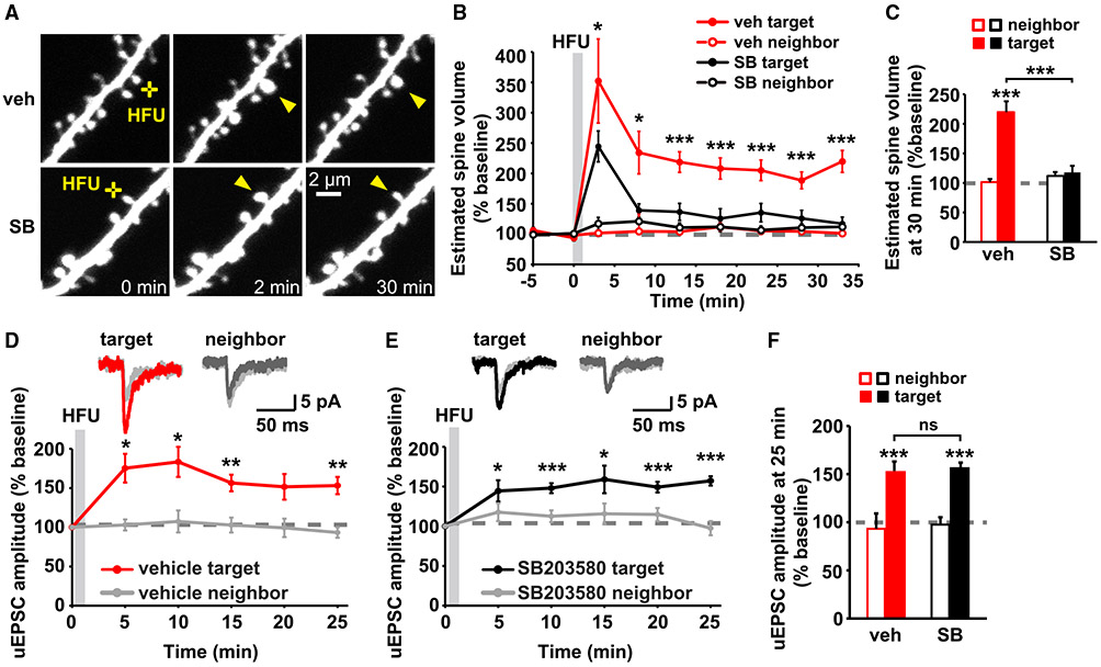
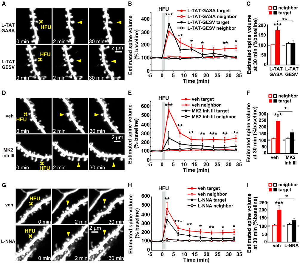
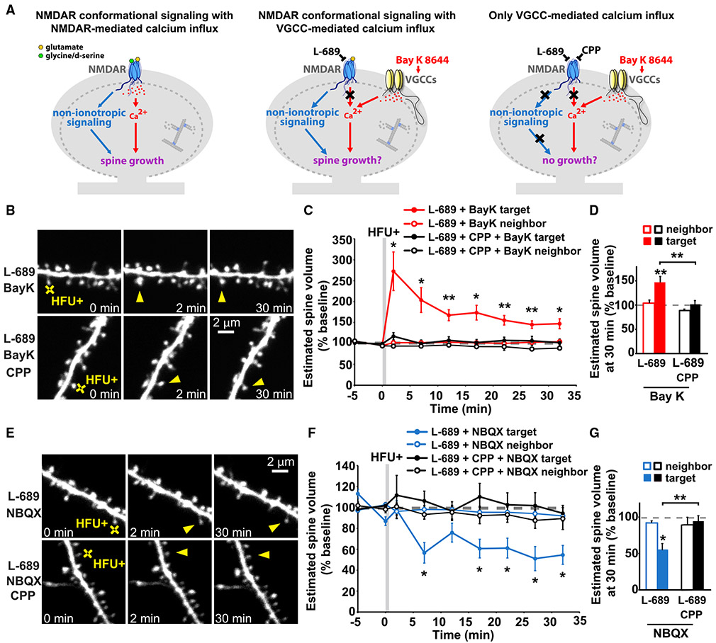
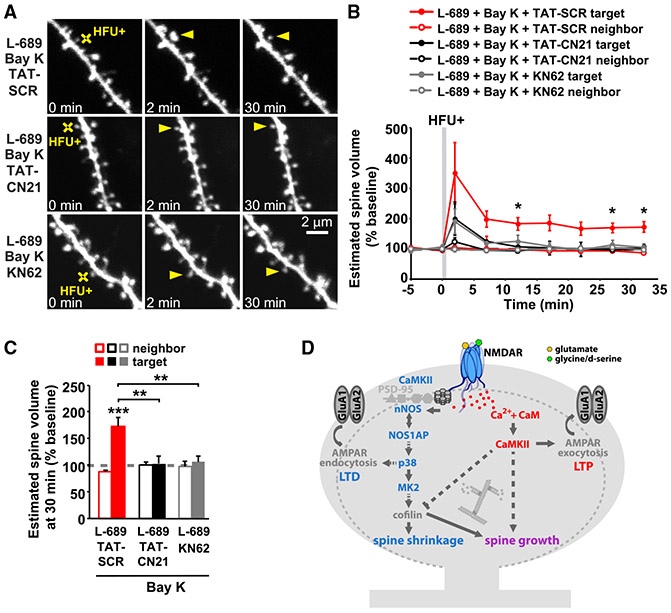
Experience-dependent refinement of neuronal connections is critically important for brain development and learning. Here, we show that ion-flow-independent NMDA receptor (NMDAR) signaling is required for the long-term dendritic spine growth that is a vital component of brain circuit plasticity. We find that inhibition of p38 mitogen-activated protein kinase (p38 MAPK), which is downstream of non-ionotropic NMDAR signaling in long-term depression (LTD) and spine shrinkage, blocks long-term potentiation (LTP)-induced spine growth but not LTP. We hypothesize that non-ionotropic NMDAR signaling drives the cytoskeletal changes that support bidirectional spine structural plasticity. Indeed, we find that key signaling components downstream of non-ionotropic NMDAR function in LTD-induced spine shrinkage are also necessary for LTP-induced spine growth. Furthermore, NMDAR conformational signaling with coincident Ca2+ influx is sufficient to drive CaMKII-dependent long-term spine growth, even when Ca2+ is artificially driven through voltage-gated Ca2+ channels. Our results support a model in which non-ionotropic NMDAR signaling gates the bidirectional spine structural changes vital for brain plasticity.

Keywords: CaMKII; LTD; LTP; NMDA receptor; dendritic spine; non-ionotropic; p38 MAPK; structural plasticity.


Conflict of interest statement

Declaration of interests The authors declare no competing interests.

Figures

Figure 1.
Figure 1.. p38 MAPK activity is required for LTP-induced spine growth, but not for synaptic strengthening
(A) Images of dendrites from DIV14-18 EGFP-expressing CA1 neurons from rat organotypic slices before and after HFU stimulation (yellow crosses) at individual dendritic spines (yellow arrowheads) with and without p38 MAPK inhibitor SB203580 (SB, 2 μM). (B and C) Inhibition of p38 MAPK with SB (black-filled circles/bar; 11 spines/11 cells) reduced HFU-induced spine growth compared with vehicle (red-filled circles/bar; 11 spines/11 cells). Volume of unstimulated neighbors (open circles/bars) was unaffected. (D and E) Top, average traces of uEPSCs from a target spine and an unstimulated neighbor on CA1 neurons from mouse organotypic slices before (gray) and 25 min after HFU stimulation during vehicle conditions (target, red; neighbor, dark gray) or in the presence of SB (target, black; neighbor, dark gray). Bottom, HFU-induced increases in uEPSC amplitude in vehicle (red; 8 spines/8 cells) were unaffected by p38 MAPK inhibition with SB (black; 9 spines/9 cells). uEPSC amplitudes of unstimulated neighboring spines (gray) were unaffected. (F) HFU induced a long-lasting uEPSC amplitude increase (red filled bar) compared with baseline, which is unaffected by p38 MAPK inhibition (black-filled bar). Two-way ANOVA with Tukey’s test used in (C) and (F) and two-way repeated-measures (RMs) ANOVA with Dunnett’s test to baseline used in (B), (D), and (E). Data are represented as means ± SEM. *p < 0.05, **p < 0.01, ***p < 0.001.
Figure 2.
Figure 2.. Non-ionotropic NMDAR signaling pathway is required for LTP-induced spine growth
(A, D, and G) Images of dendrites from EGFP-expressing CA1 neurons from rat (A and D) and mice (G) organotypic slices at DIV14-18 before and after HFU stimulation (yellow crosses) of individual spines (yellow arrowheads) in the presence of L-TAT-GESV (1 μM) and the L-TAT-GASA control peptide (1 μM), during vehicle conditions and in the presence of MK2 inhibitor III (10 μM) or the NOS inhibitor, L-NNA (100 μM). (B and C) Disruption of the NOS1AP/nNOS interaction with L-TAT-GESV (black-filled circles/bar; 9 spines/9 cells), but not application of the inactive L-TAT-GASA control peptide (red-filled circles/bar; 9 spines/9 cells) inhibited persistent spine growth after LTP induction. Volume of the unstimulated neighbors (open circles/bars) was unchanged. (E and F) Inhibition of MK2 activity (black-filled circles/bar; 11 spines/11 cells) prevented HFU-induced persistent spine enlargement (red-filled circles/bar; 12 spines/12 cells). Volume of the unstimulated neighbors did not change (open circles/bars). (H and I) Inhibition of NOS activity (black-filled circles/bar; 11 spines/11 cells) prevented HFU-induced persistent spine enlargement (red-filled circles/bar; 12 spines/12 cells). Volume of the unstimulated neighbors did not change (open circles/bars). Two-way ANOVA with Tukey’s test used in (C), (F), and (I) and two-way RMs ANOVA with Dunnett’s test to baseline used in B, E, and H. Data are represented as means ± SEM. *p < 0.05, **p < 0.01, ***p < 0.001.
Figure 3.
Figure 3.. Non-ionotropic NMDAR signaling with Ca2+-influx through voltage-gated calcium channels is sufficient to drive LTP-induced spine growth
(A) Left: proposed model in which activity-induced spine growth requires both non-ionotropic and ionotropic NMDAR signaling. Middle: schematic of experiment to test proposed model. Non-ionotropic NMDAR signaling is activated with glutamate, whereas calcium influx through the NMDAR is blocked with L-689. Instead, calcium influx is driven through VGCCs with the stronger HFU+ conditions in the presence of Bay K to favor opening of VGCCs. Right: control experiments block non-ionotropic NMDAR signaling by blocking glutamate binding to the NMDAR with CPP. (B) Images of dendrites from CA1 neurons of acute slices from P16-20 GFP-M mice before and after HFU+ stimulation (yellow crosses) of individual spines (yellow arrowheads) in the presence of L-689 (10 μM) and Bay K (10 μM) or in combination with CPP (50 μM). (C and D) HFU+ stimulation drives spine growth in the presence of Bay K, even when ion flow through the NMDAR is blocked with L-689 (red-filled circles/bar; 9 spines/9 cells), but not when non-ionotropic NMDAR signaling is blocked with CPP (black-filled circles/bar; 10 spines/10 cells). Volume of the unstimulated neighbors (open circles/bars) was unchanged. (E) Images of dendrites under experimental conditions in (B)–(D), except blocking the influx of calcium through VGCCs by removing Bay K and adding NBQX (50 μM). (F and G) HFU+ stimulation in the presence of L-689 and NBQX (blue-filled circles/bar; 7 spines/7 cells) led to spine shrinkage, which was blocked when non-ionotropic NMDAR signaling was inhibited with the presence of CPP (black-filled circles/bar; 6 spines/6 cells). Volume of the unstimulated neighbors (open circles/bars) did not change. Two-way ANOVA with Tukey’s test used in (D) and (G) and two-way RMs ANOVA with Dunnett’s test to baseline used in C and F. Data are represented as means ± SEM. *p < 0.05, **p < 0.01, ***p < 0.001. See also Figure S2.
Figure 4.
Figure 4.. CaMKII activity is required for LTP-induced spine growth, independent of calcium source
(A) Images of dendrites from CA1 neurons of acute slices from P16-20 GFP-M mice before and after HFU+ stimulation (yellow crosses) of individual spines (yellow arrowheads) in the presence of L-689 (10 μM) and Bay K (10 μM) or in combination with TAT-SCR (5 μM), TAT-CN21 (5 μM), or KN-62 (10 μM). (B and C) HFU+-induced spine growth in the presence of Bay K and L-689 is blocked by TAT-CN21 peptide (black-filled circles/bar; 6 spines/ 6 cells) or KN-62 (gray-filled circles/bars; 6 spines/6 cells), but not in the presence of control TAT-SCR peptide (red-filled circles/bar; 6 spines/ 6 cells). Volume of the unstimulated neighbors (open circles/bars) was unchanged. Two-way ANOVA with Tukey’s test used in (C) and two-way RMs ANOVA with Dunnett’s test to baseline used in B. Data are represented as means ± SEM. *p < 0.05, **p < 0.01, ***p < 0.001. (D) Proposed model. Plasticity-inducing glutamatergic stimulation activates non-ionotropic NMDAR signaling, driving cofilin-dependent severing of the actin cytoskeleton, which, in the absence of strong Ca2+ influx, leads to spine shrinkage. On the other hand, during the strong Ca2+ influx associated with LTP induction, severed actin filaments serve as new starting points for actin filament nucleation and branching by the Ca2+- and CaMKII-dependent actin-modifying proteins to expand the F-actin cytoskeleton and drive spine growth.

Similar articles

Cited by

References

    1. Anderson DR, Meyers MJ, Vernier WF, Mahoney MW, Kurumbail RG, Caspers N, Poda GI, Schindler JF, Reitz DB, and Mourey RJ (2007). Pyrrolopyridine inhibitors of mitogen-activated protein kinase-activated protein kinase 2 (MK-2). J. Med. Chem 50, 2647–2654. - PubMed
    1. Aow J, Dore K, and Malinow R (2015). Conformational signaling required for synaptic plasticity by the NMDA receptor complex. Proc. Natl. Acad. Sci. USA 112, 14711–14716. - PMC - PubMed
    1. Barria A, and Malinow R (2005). NMDA receptor subunit composition controls synaptic plasticity by regulating binding to CaMKII. Neuron 48, 289–301. - PubMed
    1. Bayer KU, and Schulman H (2019). CaM Kinase: Still Inspiring at 40. Neuron 103, 380–394. - PMC - PubMed
    1. Birnbaum JH, Bali J, Rajendran L, Nitsch RM, and Tackenberg C (2015). Calcium flux-independent NMDA receptor activity is required for Aβ oligomer-induced synaptic loss. Cell Death Dis. 6, e1791. - PMC - PubMed

Publication types

MeSH terms

Substances