. 2021 Jan;48(1):84-90.
doi: 10.5999/aps.2020.00969. Epub 2021 Jan 15.

Vascularized bone grafts for post-traumatic defects in the upper extremity

Affiliations

Vascularized bone grafts for post-traumatic defects in the upper extremity

Giovanna Petrella et al. Arch Plast Surg. 2021 Jan.

Abstract

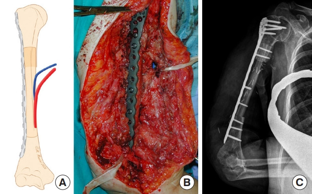
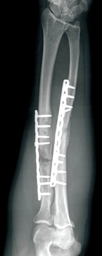
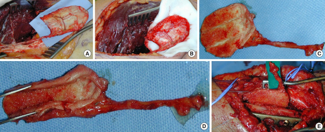
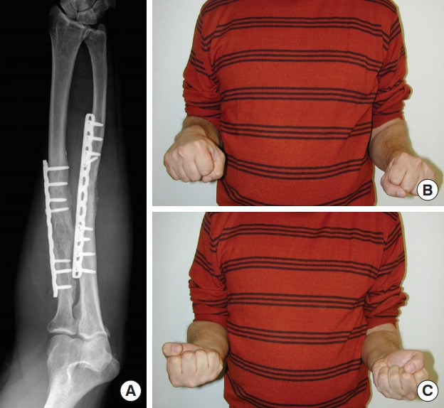
Vascularized bone grafts (VBGs) are widely employed to reconstruct upper extremity bone defects. Conventional bone grafting is generally used to treat defects smaller than 5-6 cm, when tissue vascularization is adequate and there is no infection risk. Vascularized fibular grafts (VFGs) are mainly used in the humerus, radius or ulna in cases of persistent non-union where traditional bone grafting has failed or for bone defects larger than 6 cm. Furthermore, VFGs are considered to be the standard treatment for large bone defects located in the radius, ulna and humerus and enable the reconstruction of soft-tissue loss, as VFGs can be harvested as osteocutaneous flaps. VBGs enable one-stage surgical reconstruction and are highly infection-resistant because of their autonomous vascularization. A vascularized medial femoral condyle (VFMC) free flap can be used to treat small defects and non-unions in the upper extremity. Relative contraindications to these procedures are diabetes, immunosuppression, chronic infections, alcohol, tobacco, drug abuse and obesity. The aim of our study was to illustrate the use of VFGs to treat large post-traumatic bone defects and osteomyelitis located in the upper extremity. Moreover, the use of VFMC autografts is presented.

Keywords: Bone; Cortico-periosteal; Fibula; Graft; Microsurgery.


Conflict of interest statement

Conflict of interest

No potential conflict of interest relevant to this article was reported.

Figures

Fig. 1.
Fig. 1.. Necrotic radius, infected bone debridement
Case 1. The radius had been operated on four times within 3 years, with complete resorption of the conventional bone graft (A). After removing the plate (B), extensive debridement and excision of all necrotic bone were performed (C), creating a segmental defect requiring reconstruction (D).
Fig. 2.
Fig. 2.. Free fibula flap with periosteal sparing
Case 1. (A) A sleeve of vascularized periosteum is retained with a vascularized fibular graft. (B) Plate was fixed using a single locking compression plate. The sleeve of vascularized periosteum was cuffed around the recipient bone (white arrows) to enhance healing at the junction site.
Fig. 3.
Fig. 3.. Outcomes of case 1
(A) Radiographic evaluation after 6 months. (B, C) Clinical evaluation: pronosupination is maintained (right hand).
Fig. 4.
Fig. 4.. Humerus non-union
(A) Recalcitrant non-union of the humerus, with multiple failed operations. (B) Reconstruction was performed with a vascularized fibular graft (VFG). Internal fixation was achieved with a locking compression plate. (C) Twelve months after surgery, X-rays revealed union and relevant hypertrophy of the VFG.
Fig. 5.
Fig. 5.. Step-cut osteotomy
(A) Schematic representation of step-cut osteotomy for ulna reconstruction. (B) X-ray examination showing postoperative outcomes.
Fig. 6.
Fig. 6.. Transverse osteotomy
(A) Schematic representation of transverse osteotomy for radius reconstruction. (B) Preplating the vascularized fibular graft. (C) Intraoperative view after osteosynthesis with a locking compression plate. (D) X-ray examination showing postoperative outcomes.
Fig. 7.
Fig. 7.. Intramedullary press-fit insetting
(A) Schematic representation of an intramedullary press-fit for humerus reconstruction. (B) Intraoperative result after osteosynthesis with a long bridging plate. (C) X-ray examination showing postoperative outcomes.
Fig. 8.
Fig. 8.. Staged repair in forearm infection
Case 2. (A, B) Open ulnar fracture treated 4 months prior with a plate, with clinical and radiological features of severe infection in the forearm. (C, D) After debridement, a cement spacer was applied. (E) Three months after, vascularized free graft reconstruction was planned. (F) Radiological result of the reconstructed ulna, 12 months after surgery.
Fig. 9.
Fig. 9.. Recalcitrant radius non-union
Case 3. Radiographic appearance.
Fig. 10.
Fig. 10.. Corticoperiosteal flap
Case 3. (A) A thin corticoperiosteal flap is harvested from the medial surface of the supracondylar region. The graft is cut with the aid of a chisel (proceeding from distal to proximal to avoid vessel injury). (B) Good vascular supply of the flap after releasing the tourniquet. (C) The medial femoral condylar periosteal flap is smaller and more flexible and may be easily tailored to irregular defects. (D) Subperiosteal resection of two or three 2-mm-wide strips of cortical layer allows easier flap wrapping around the bone graft. (E) The flap is placed around the radius and fixed with wire or nonabsorbable suture, and microvascular anastomosis is subsequently performed to the adjacent vessels.
Fig. 11.
Fig. 11.. Outcomes of case 3
Radiographic (A) and clinical (B, C) outcomes at 4 months of follow-up.

Similar articles

Cited by

References

    1. Klifto CS, Gandi SD, Sapienza A. Bone graft options in upper-extremity surgery. J Hand Surg Am. 2018;43:755–61. - PubMed
    1. Khan SN, Cammisa FP, Jr, Sandhu HS, et al. The biology of bone grafting. J Am Acad Orthop Surg. 2005;13:77–86. - PubMed
    1. Taylor GI, Miller GD, Ham FJ. The free vascularized bone graft: a clinical extension of microvascular techniques. Plast Reconstr Surg. 1975;55:533–44. - PubMed
    1. Adani R, Delcroix L, Innocenti M, et al. Reconstruction of large posttraumatic skeletal defects of the forearm by vascularized free fibular graft. Microsurgery. 2004;24:423–9. - PubMed
    1. Safoury Y. Free vascularized fibula for the treatment of traumatic bone defects and nonunion of the forearm bones. J Hand Surg Br. 2005;30:67–72. - PubMed