

A Novel Role for the DNA Repair Enzyme 8-Oxoguanine DNA Glycosylase in Adipogenesis
- PMID: 33503804
- PMCID: PMC7865743
- DOI: 10.3390/ijms22031152
A Novel Role for the DNA Repair Enzyme 8-Oxoguanine DNA Glycosylase in Adipogenesis
Abstract
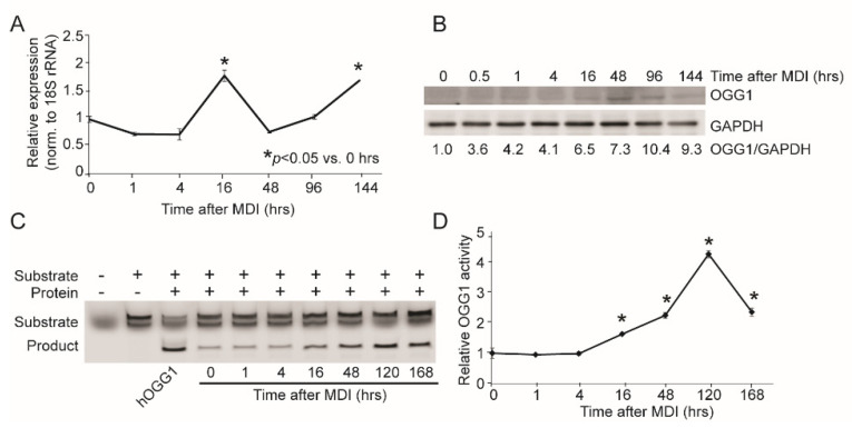
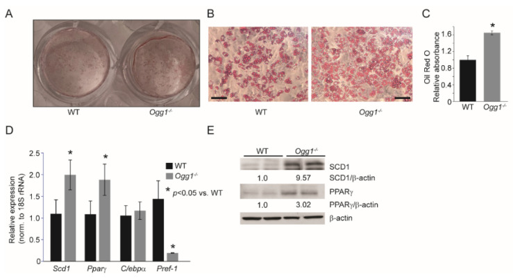
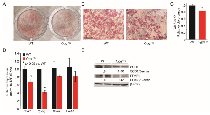
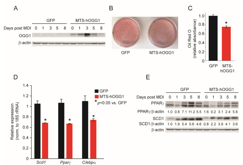
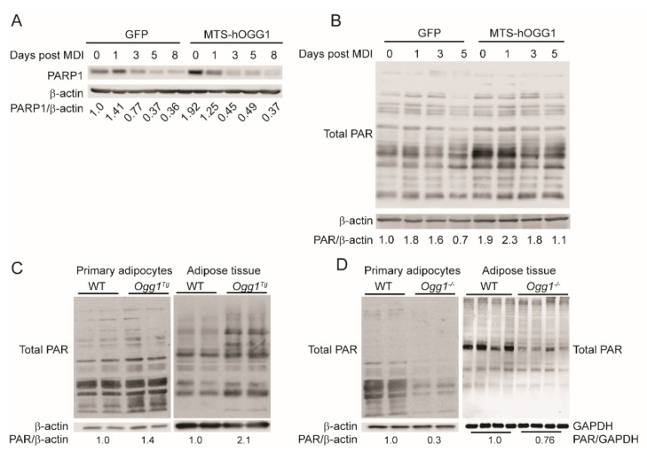
Cells sustain constant oxidative stress from both exogenous and endogenous sources. When unmitigated by antioxidant defenses, reactive oxygen species damage cellular macromolecules, including DNA. Oxidative lesions in both nuclear and mitochondrial DNA are repaired via the base excision repair (BER) pathway, initiated by DNA glycosylases. We have previously demonstrated that the BER glycosylase 8-oxoguanine DNA glycosylase (OGG1) plays a novel role in body weight maintenance and regulation of adiposity. Specifically, mice lacking OGG1 (Ogg1-/-) are prone to increased fat accumulation with age and consumption of hypercaloric diets. Conversely, transgenic animals with mitochondrially-targeted overexpression of OGG1 (Ogg1Tg) are resistant to age- and diet-induced obesity. Given these phenotypes of altered adiposity in the context of OGG1 genotype, we sought to determine if OGG1 plays a cell-intrinsic role in adipocyte maturation and lipid accumulation. Here, we report that preadipocytes from Ogg1-/- mice differentiate more efficiently and accumulate more lipids than those from wild-type animals. Conversely, OGG1 overexpression significantly blunts adipogenic differentiation and lipid accretion in both pre-adipocytes from Ogg1Tg mice, as well as in 3T3-L1 cells with adenovirus-mediated OGG1 overexpression. Mechanistically, changes in adipogenesis are accompanied by significant alterations in cellular PARylation, corresponding with OGG1 genotype. Specifically, deletion of OGG1 reduces protein PARylation, concomitant with increased adipogenic differentiation, while OGG1 overexpression significantly increases PARylation and blunts adipogenesis. Collectively, these data indicate a novel role for OGG1 in modulating adipocyte differentiation and lipid accretion. These findings have important implications to our knowledge of the fundamental process of adipocyte differentiation, as well as to our understanding of lipid-related diseases such as obesity.
Keywords: DNA repair; adipocyte differentiation; base excision repair; lipid accretion; obesity.
Conflict of interest statement
The authors declare no conflict of interest.
Figures
Similar articles
-
8-oxoguanine DNA glycosylase (OGG1) deficiency elicits coordinated changes in lipid and mitochondrial metabolism in muscle.PLoS One. 2017 Jul 20;12(7):e0181687. doi: 10.1371/journal.pone.0181687. eCollection 2017. PLoS One. 2017. PMID: 28727777 Free PMC article.
-
Activity of OGG1 variants in the repair of pro-oxidant-induced 8-oxo-2'-deoxyguanosine.DNA Repair (Amst). 2006 Nov 8;5(11):1337-45. doi: 10.1016/j.dnarep.2006.06.001. Epub 2006 Jul 24. DNA Repair (Amst). 2006. PMID: 16861056
-
Whole transcriptome analysis reveals a role for OGG1-initiated DNA repair signaling in airway remodeling.Free Radic Biol Med. 2015 Dec;89:20-33. doi: 10.1016/j.freeradbiomed.2015.07.007. Epub 2015 Jul 15. Free Radic Biol Med. 2015. PMID: 26187872 Free PMC article.
-
Repair of 8-oxo-7,8-dihydroguanine in prokaryotic and eukaryotic cells: Properties and biological roles of the Fpg and OGG1 DNA N-glycosylases.Free Radic Biol Med. 2017 Jun;107:179-201. doi: 10.1016/j.freeradbiomed.2016.11.042. Epub 2016 Nov 27. Free Radic Biol Med. 2017. PMID: 27903453 Review.
-
Lost in the Crowd: How Does Human 8-Oxoguanine DNA Glycosylase 1 (OGG1) Find 8-Oxoguanine in the Genome?Int J Mol Sci. 2020 Nov 7;21(21):8360. doi: 10.3390/ijms21218360. Int J Mol Sci. 2020. PMID: 33171795 Free PMC article. Review.
Cited by
-
Chemical Switching: A Concept Inspired by Strategies from Biocatalysis and Organocatalysis.Chembiochem. 2025 Jun 3;26(11):e202500220. doi: 10.1002/cbic.202500220. Epub 2025 May 26. Chembiochem. 2025. PMID: 40417833 Free PMC article. Review.
-
Maternal Transmission of Human OGG1 Protects Mice Against Genetically- and Diet-Induced Obesity Through Increased Tissue Mitochondrial Content.Front Cell Dev Biol. 2021 Sep 15;9:718962. doi: 10.3389/fcell.2021.718962. eCollection 2021. Front Cell Dev Biol. 2021. PMID: 34604220 Free PMC article.
-
Organocatalytic Switches of DNA Glycosylase OGG1 Catalyze a Highly Efficient AP-Lyase Function.Chemistry. 2025 Jun 12;31(33):e202500382. doi: 10.1002/chem.202500382. Epub 2025 May 15. Chemistry. 2025. PMID: 40294343 Free PMC article.
-
8-Oxoguanine DNA Glycosylase 1 Upregulation as a Risk Factor for Obesity and Colorectal Cancer.Int J Mol Sci. 2023 Mar 13;24(6):5488. doi: 10.3390/ijms24065488. Int J Mol Sci. 2023. PMID: 36982562 Free PMC article.
-
DNA Mutagenicity of Hydroxyhydroquinone in Roasted Coffee Products and Its Suppression by Chlorogenic Acid, a Coffee Polyphenol, in Oxidative-Damage-Sensitive SAMP8 Mice.Int J Mol Sci. 2024 Jan 5;25(2):720. doi: 10.3390/ijms25020720. Int J Mol Sci. 2024. PMID: 38255794 Free PMC article.
References
-
- Abolhassani N., Leon J., Sheng Z., Oka S., Hamasaki H., Iwaki T., Nakabeppu Y. Molecular pathophysiology of impaired glucose metabolism, mitochondrial dysfunction, and oxidative DNA damage in Alzheimer’s disease brain. Mech. Ageing Dev. 2016;161:95–104. doi: 10.1016/j.mad.2016.05.005. - DOI - PubMed
-
- Audebert M., Chevillard S., Levalois C., Gyapay G., Vieillefond A., Klijanienko J., Vielh P., El Naggar A.K., Oudard S., Boiteux S., et al. Alterations of the DNA repair gene OGG1 in human clear cell carcinomas of the kidney. Cancer Res. 2000;60:4740–4744. - PubMed
MeSH terms
Substances
Grants and funding
LinkOut - more resources
Full Text Sources
Other Literature Sources
Molecular Biology Databases
Research Materials
Miscellaneous
