. 2021 Jan 25;26(3):606.
doi: 10.3390/molecules26030606.

JAK2 Inhibition by Fedratinib Reduces Osteoblast Differentiation and Mineralisation of Human Mesenchymal Stem Cells

Affiliations

JAK2 Inhibition by Fedratinib Reduces Osteoblast Differentiation and Mineralisation of Human Mesenchymal Stem Cells

Nihal AlMuraikhi et al. Molecules. .

Abstract

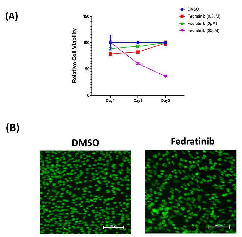
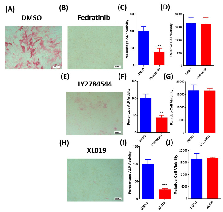
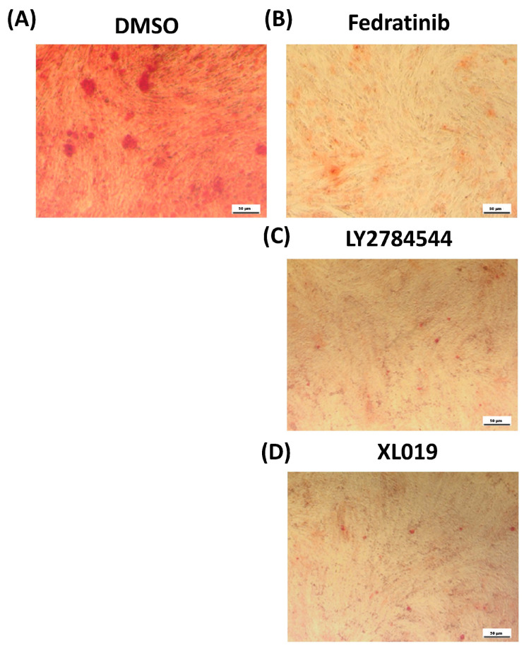
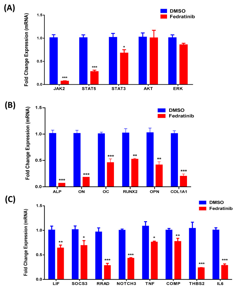
Several signalling pathways, including the JAK/STAT signalling pathway, have been identified to regulate the differentiation of human bone marrow skeletal (mesenchymal) stem cells (hBMSCs) into bone-forming osteoblasts. Members of the JAK family mediate the intracellular signalling of various of cytokines and growth factors, leading to the regulation of cell proliferation and differentiation into bone-forming osteoblastic cells. Inhibition of JAK2 leads to decoupling of its downstream mediator, STAT3, and the subsequent inhibition of JAK/STAT signalling. However, the crucial role of JAK2 in hBMSCs biology has not been studied in detail. A JAK2 inhibitor, Fedratinib, was identified during a chemical biology screen of a small molecule library for effects on the osteoblastic differentiation of hMSC-TERT cells. Alkaline phosphatase activity and staining assays were conducted as indicators of osteoblastic differentiation, while Alizarin red staining was used as an indicator of in vitro mineralised matrix formation. Changes in gene expression were assessed using quantitative real-time polymerase chain reaction. Fedratinib exerted significant inhibitory effects on the osteoblastic differentiation of hMSC-TERT cells, as demonstrated by reduced ALP activity, in vitro mineralised matrix formation and downregulation of osteoblast-related gene expression, including ALP, ON, OC, RUNX2, OPN, and COL1A1. To identify the underlying molecular mechanisms, we examined the effects of Fedratinib on a molecular signature of several target genes known to affect hMSC-TERT differentiation into osteoblasts. Fedratinib inhibited the expression of LIF, SOCS3, RRAD, NOTCH3, TNF, COMP, THBS2, and IL6, which are associated with various signalling pathways, including TGFβ signalling, insulin signalling, focal adhesion, Notch Signalling, IL-6 signalling, endochondral ossification, TNF-α, and cytokines and inflammatory response. We identified a JAK2 inhibitor (Fedratinib) as a powerful inhibitor of the osteoblastic differentiation of hMSC-TERT cells, which may be useful as a therapeutic option for treating conditions associated with ectopic bone formation or osteosclerotic metastases.

Keywords: Fedratinib; JAK/STAT signalling pathway; JAK2 inhibition; hMSC-TERT; in vitro mineralisation; osteoblast differentiation.


Conflict of interest statement

The authors declare no conflict of interest.

Figures

Figure 1
Figure 1
Effects of Fedratinib treatment on the number of hMSC-TERT cells. (A) Dose-response curve of hMSC-TERT cells to different doses of Fedratinib, as indicated in the graph, vs. DMSO-treated control cells as measured by cell number assay over 3 days. (B) Representative fluorescence images of Fedratinib-treated hMSC-TERT cells (3.0 µM) vs. DMSO-treated control cells on day 3 of exposure. Photomicrograph magnification, 20×. Cells were stained with AO/EtBr to detect apoptotic (cells with green condensed chromatin) and necrotic (red) cells. DMSO: dimethyl sulfoxide.
Figure 2
Figure 2
Effects of JAK2 inhibitors on the osteoblastic differentiation of hMSC-TERT cells. (A) Representative ALP staining of DMSO-treated control cells on day 10 post-osteoblastic differentiation. Photomicrograph magnification, 10×. (B) Representative ALP staining of Fedratinib-treated hMSC-TERT cells (3.0 µM) on day 10 post-osteoblastic differentiation. Photomicrograph magnification, 10×. (C) Quantification of ALP activity in Fedratinib-treated hMSC-TERT cells (3.0 µM) vs. DMSO-treated control cells on day 10 post-osteoblastic differentiation. Data are presented as the mean percentage of ALP activity ± SEM (n = 20). (D) Assay of cell viability using alamarBlue in Fedratinib-treated hMSC-TERT cells (3.0 µM) vs. DMSO-treated control cells on day 10 post-osteoblastic differentiation. Data are presented as the mean ± SEM (n = 20). (E) Representative ALP staining of LY2784544-treated hMSC-TERT cells (3.0 µM) on day 10 post-osteoblastic differentiation. Photomicrograph magnification, 10×. (F) Quantification of ALP activity in LY2784544-treated hMSC-TERT cells (3.0 µM) vs. DMSO-treated control cells on day 10 post-osteoblastic differentiation. Data are presented as the mean percentage of ALP activity ± SEM (n = 20). (G) Assay of cell viability using alamarBlue assay in LY2784544-treated hMSC-TERT cells (3.0 µM) vs. DMSO-treated control cells on day 10 post-osteoblastic differentiation. Data are presented as the mean ± SEM (n = 20). (H) Representative ALP staining of XL019-treated hMSC-TERT cells (3.0 µM) on day 10 post-osteoblastic differentiation. Photomicrograph magnification, 10×. (I) Quantification of ALP activity in XL019-treated hMSC-TERT cells (3.0 µM) vs. DMSO-treated control cells on day 10 post-osteoblastic differentiation. Data are presented as the mean percentage ALP activity ± SEM (n = 20). (J) Assay of cell viability using alamarBlue assay in XL019-treated hMSC-TERT cells (3.0 µM) vs. DMSO-treated control cells on day 10 post-osteoblastic differentiation. Data are presented as the mean ± SEM (n = 20). ALP, alkaline phosphatase; DMSO, dimethyl sulfoxide. ** p < 0.005; *** p < 0.0005.
Figure 3
Figure 3
Effects of JAK2 inhibitors on the mineralisation of hMSC-TERT cells. (A) Cytochemical staining of mineralised matrix formation using Alizarin red on day 21 post-osteoblastic differentiaTable 21. post-osteoblastic differentiation in Fedratinib-treated hMSC-TERT cells (3.0 µM). (C) Cytochemical staining of mineralised matrix formation using Alizarin red on day 21 post-osteoblastic differentiation in LY2784544-treated hMSC-TERT cells (3.0 µM). (D) Cytochemical staining of mineralised matrix formation using Alizarin red on day 21 post-osteoblastic differentiation in XL019-treated hMSC-TERT cells (3.0 µM). Photomicrograph magnification, 10×.
Figure 4
Figure 4
Fedratinib downregulates multiple osteoblast-associated genes. (A) Quantitative RT-PCR analysis of the expression of JAK2, STAT5, STAT3, AKT, and ERK in hMSC-TERT cells after 24 h of osteoblastic differentiation in the absence (blue) or presence (red) of Fedratinib (3.0 µM). (B) Quantitative RT-PCR analysis of the expression of ALP, ON, OC, RUNX2, OPN, and COL141 in hMSC-TERT cells on day 10 post osteoblastic differentiation in the absence (blue) or presence (red) of Fedratinib (3.0 µM). Alkaline phosphatase (ALP); Osteonectin (ON); Osteocalcin (OC); Runt-related transcription factor 2 (RUNX2); Osteopontin (OPN); Collagen type I alpha 1 (COL1A1); Dimethyl sulfoxide (DMSO). (C) Quantitative RT-PCR analysis of a selected panel of osteoblast differentiation-associated genes in Fedratinib-treated hMSC-TERT cells vs. DMSO-treated control cells using qRT-PCR on day 10 post osteoblastic differentiation in the absence (blue) or presence (red) of Fedratinib (3.0 µM). Gene expression was normalised to that of β-actin. Data are presented as the mean fold change ± SEM (n = 6) in replicates from two independent experiments; * p < 0.05; ** p < 0.005; *** p < 0.0005.

References

    1. Darvin P., Joung Y.H., Yang Y.M. JAK2-STAT5B pathway and osteoblast differentiation. JAKSTAT. 2013;2:e24931. doi: 10.4161/jkst.24931. - DOI - PMC - PubMed
    1. Adam S., Simon N., Steffen U., Andes F.T., Scholtysek C., Muller D.I.H., Weidner D., Andreev D., Kleyer A., Culemann S., et al. JAK inhibition increases bone mass in steady-state conditions and ameliorates pathological bone loss by stimulating osteoblast function. Sci. Transl. Med. 2020;12 doi: 10.1126/scitranslmed.aay4447. - DOI - PubMed
    1. Li J. JAK-STAT and bone metabolism. JAKSTAT. 2013;2:e23930. doi: 10.4161/jkst.23930. - DOI - PMC - PubMed
    1. Jatiani S.S., Baker S.J., Silverman L.R., Reddy E.P. Jak/STAT pathways in cytokine signaling and myeloproliferative disorders: Approaches for targeted therapies. Genes Cancer. 2010;1:979–993. doi: 10.1177/1947601910397187. - DOI - PMC - PubMed
    1. Ji J., Wu Y., Meng Y., Zhang L., Feng G., Xia Y., Xue W., Zhao S., Gu Z., Shao X. JAK-STAT signaling mediates the senescence of bone marrow-mesenchymal stem cells from systemic lupus erythematosus patients. Acta Biochim. Biophys. Sin. 2017;49:208–215. doi: 10.1093/abbs/gmw134. - DOI - PubMed

MeSH terms

LinkOut - more resources