. 2021 Jan 25;22(3):1161.
doi: 10.3390/ijms22031161.

Cardiomyocytes Derived from Induced Pluripotent Stem Cells as a Disease Model for Propionic Acidemia

Affiliations

Cardiomyocytes Derived from Induced Pluripotent Stem Cells as a Disease Model for Propionic Acidemia

Esmeralda Alonso-Barroso et al. Int J Mol Sci. .

Abstract

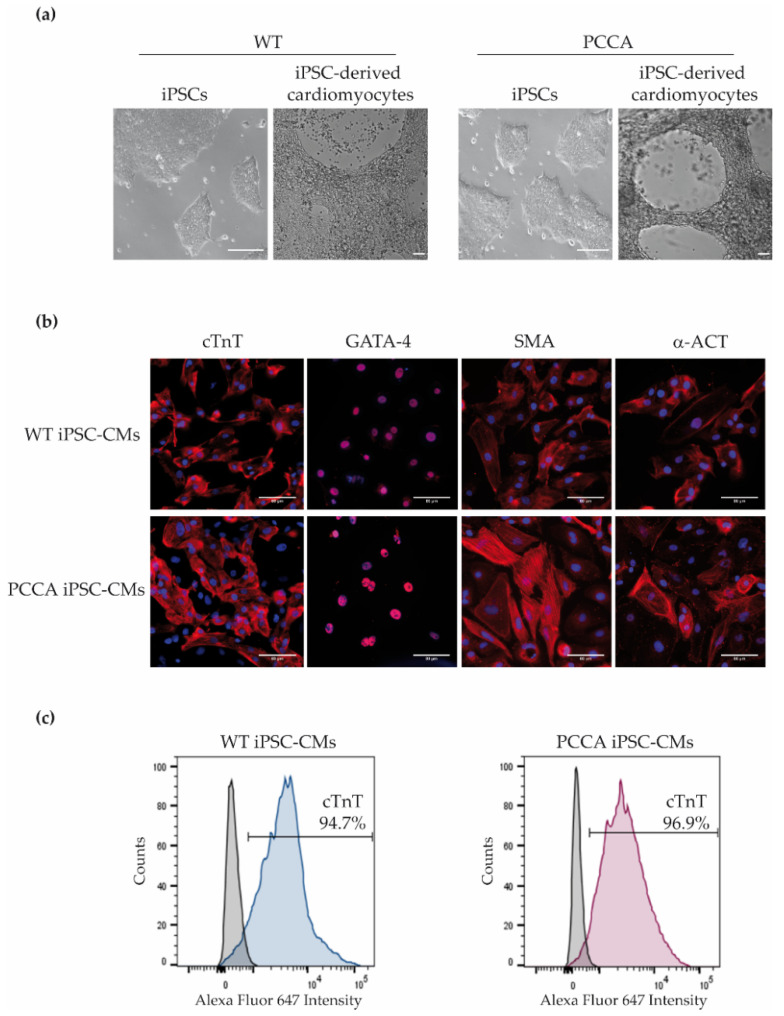
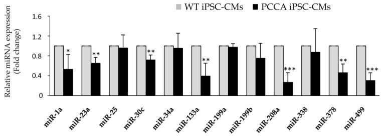
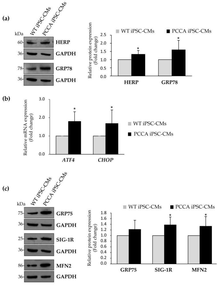
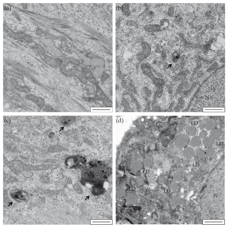
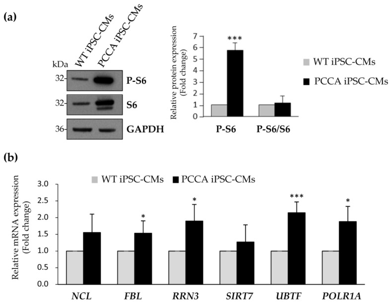
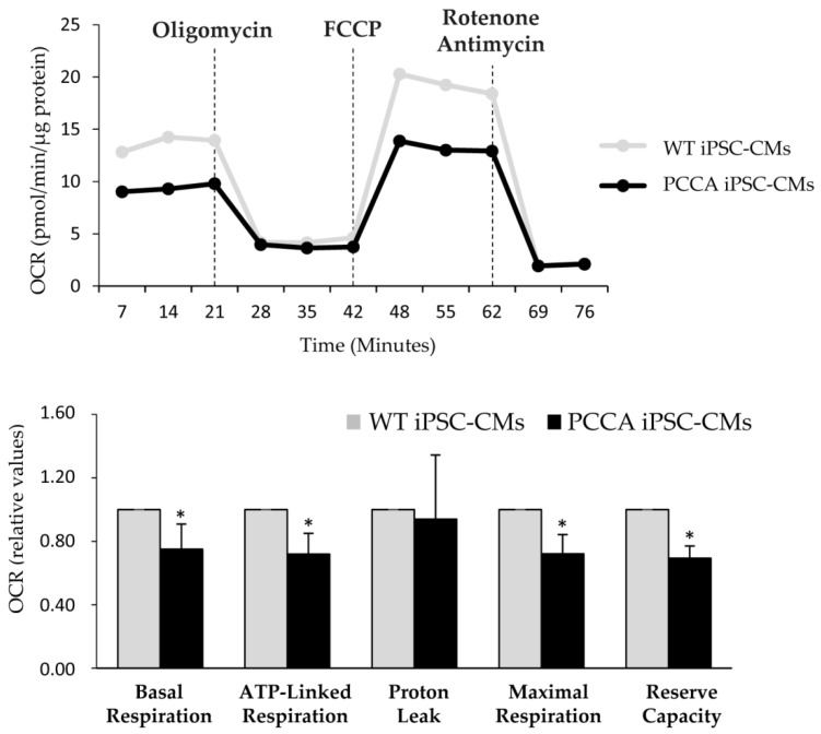
Propionic acidemia (PA), one of the most frequent life-threatening organic acidemias, is caused by mutations in either the PCCA or PCCB genes encoding both subunits of the mitochondrial propionyl-CoA carboxylase (PCC) enzyme. Cardiac alterations (hypertrophy, dilated cardiomyopathy, long QT) are one of the major causes of mortality in patients surviving the neonatal period. To overcome limitations of current cellular models of PA, we generated induced pluripotent stem cells (iPSCs) from a PA patient with defects in the PCCA gene, and successfully differentiated them into cardiomyocytes. PCCA iPSC-derived cardiomyocytes exhibited reduced oxygen consumption, an accumulation of residual bodies and lipid droplets, and increased ribosomal biogenesis. Furthermore, we found increased protein levels of HERP, GRP78, GRP75, SIG-1R and MFN2, suggesting endoplasmic reticulum stress and calcium perturbations in these cells. We also analyzed a series of heart-enriched miRNAs previously found deregulated in the heart tissue of a PA murine model and confirmed their altered expression. Our novel results show that PA iPSC-cardiomyocytes represent a promising model for investigating the pathological mechanisms underlying PA cardiomyopathies, also serving as an ex vivo platform for therapeutic evaluation.

Keywords: cardiac dysfunction; disease model; iPSC; iPSC-derived cardiomyocytes; propionic acidemia.


Conflict of interest statement

The authors declare no conflict of interest.

Figures

Figure 1
Figure 1
Phase-contrast pictures of iPSCs and differentiated cardiomyocytes and expression of cardiac markers in wild-type (WT) and PCCA iPSC-derived cardiomyocytes (iPSC-CMs). (a) Phase-contrast images of iPSC and iPSC-derived cardiomyocytes of WT and PCCA; scale bar: 100 µm. (b) Immunofluorescence analysis for cardiac troponin T (cTnT), GATA-4, α-smooth muscle actin (SMA) and α-actinin (α-ACT) in iPSC-derived cardiomyocytes; scale bar: 80 µm. (c) Flow cytometry analysis for cTnT cardiac marker. A representative experiment for cTnT expression is shown.
Figure 2
Figure 2
Analysis of miRNA expression in WT and PCCA iPSC-CMs. Relative expression levels of miR-1a, miR-23a, miR-25, miR-30c, miR-34a, miR-133a, miR-199a, miR-199b, miR-208a, miR-338, miR-378 and miR-499 are evaluated by qRT-PCR in iPSC-derived cardiomyocytes. Data represents mean ± standard deviation of three independent cardiomyocyte differentiation triplicates at least. Statistical significance is determined by the Student’s t-test. * p < 0.05; ** p < 0.01; *** p < 0.001.
Figure 3
Figure 3
Evaluation of protein and mRNA levels involved in the unfolded protein response (UPR) and mitochondria-associated membranes (MAMs) in WT and PCCA iPSC-CMs. (a,b) Expression analysis of several proteins involved in the UPR by western blot (a) or by qRT-PCR (b). (c) Analysis of protein levels of MAMs proteins by western blot. In panels (a,c), representative blots and the corresponding quantification of proteins by laser densitometry are shown as the mean ± standard deviation of at least three experiments. In each blot, GADPH is used as a loading control. In (b), data represents the mean ± standard deviation of at least three independent cardiomyocyte differentiation triplicates. Statistical significance is determined by Student’s t-test. * p < 0.05.
Figure 4
Figure 4
Electron microscopy of iPSC-derived cardiomyocytes. Representative images are shown of WT-iPSC-CMs (a,b) and PCCA iPSC-CMs (c,d) at 5000× magnification. Black arrows show degradation vesicles (b,c). LD: lipid droplets (d). N: cell nucleus (b,d). Scale bar: 1 µm.
Figure 5
Figure 5
Analysis of expression levels of proteins involved in ribosomal biogenesis. (a) Representative blot of the analysis of S6 ribosomal protein and its phosphorylated form. GADPH is used as loading control. The corresponding quantification by laser densitometry is shown as the mean ± standard deviation of at least three experiments. (b) Relative mRNA expression of NCL, FBL, RRN3, SIRT7, UBTF and POLR1A genes by qRT-PCR. Data represents the mean ± standard deviation of three independent biological triplicates. Statistical significance is determined by Student’s t-test. * p < 0.05; *** p < 0.001.
Figure 6
Figure 6
Bioenergetic profile of WT and PCCA iPSC-CMs. Representative profile of basal oxygen consumption rate (OCR) in WT and PCCA iPSC-CMs, and after the addition of oligomycin, FCCP, rotenone and antimycin A. Relative values of OCR are shown as the mean ± standard deviation of three to five wells from three independent cardiomyocyte differentiations. Statistical significance is determined by Student’s t-test. * p < 0.05.

Similar articles

Cited by

References

    1. Fenton W.A., Gravel R.A., Rosenberg L.E. Disorders of propionate and methylmalonate metabolism. In: Scriver C.R., Beaudet A.L., Sly W., Valle D., editors. The Metabolic and Molecular Bases of Inherited Disease. 8th ed. McGraw-Hill; New York, NY, USA: 2001. pp. 2165–2190.
    1. Baumgartner D., Scholl-Burgi S., Sass J.O., Sperl W., Schweigmann U., Stein J.I., Karall D. Prolonged QTc intervals and decreased left ventricular contractility in patients with propionic acidemia. J. Pediatr. 2007;150:192–197. doi: 10.1016/j.jpeds.2006.11.043. - DOI - PubMed
    1. Pena L., Franks J., Chapman K.A., Gropman A., Mew N.A., Chakrapani A., Island E., MacLeod E., Matern D., Smith B., et al. Natural history of propionic acidemia. Mol. Genet. Metab. 2012;105:5–9. doi: 10.1016/j.ymgme.2011.09.022. - DOI - PubMed
    1. Romano S., Valayannopoulos V., Touati G., Jais J.P., Rabier D., de Keyzer Y., Bonnet D., de Lonlay P. Cardiomyopathies in propionic aciduria are reversible after liver transplantation. J. Pediatr. 2010;156:128–134. doi: 10.1016/j.jpeds.2009.07.002. - DOI - PubMed
    1. Guenzel A.J., Hofherr S.E., Hillestad M., Barry M., Weaver E., Venezia S., Kraus J.P., Matern D., Barry M.A. Generation of a hypomorphic model of propionic acidemia amenable to gene therapy testing. Mol. Ther. 2013;21:1316–1323. doi: 10.1038/mt.2013.68. - DOI - PMC - PubMed

MeSH terms