. 2021 Jan 25;11(2):153.
doi: 10.3390/biom11020153.

Reconstitution of Human Necrosome Interactions in Saccharomyces cerevisiae

Affiliations

Reconstitution of Human Necrosome Interactions in Saccharomyces cerevisiae

Y Ji et al. Biomolecules. .

Abstract

The necrosome is a large-molecular-weight complex in which the terminal effector of the necroptotic pathway, Mixed Lineage Kinase Domain-Like protein (MLKL), is activated to induce necroptotic cell death. The precise mechanism of MLKL activation by the upstream kinase, Receptor Interacting Serine/Threonine Protein Kinase 3 (RIPK3) and the role of Receptor Interacting Serine/Threonine Protein Kinase 1 (RIPK1) in mediating MLKL activation remain incompletely understood. Here, we reconstituted human necrosome interactions in yeast by inducible expression of these necrosome effectors. Functional interactions were reflected by the detection of phosphorylated MLKL, plasma membrane permeabilization, and reduced proliferative potential. Following overexpression of human necrosome effectors in yeast, MLKL aggregated in the periphery of the cell, permeabilized the plasma membrane and compromised clonogenic potential. RIPK1 had little impact on RIPK3/MLKL-mediated yeast lethality; however, it exacerbated the toxicity provoked by co-expression of MLKL with a RIPK3 variant bearing a mutated RHIM-domain. Small molecule necroptotic inhibitors necrostatin-1 and TC13172, and viral inhibitors M45 (residues 1-90) and BAV_Rmil, abated the yeast toxicity triggered by the reconstituted necrosome. This yeast model provides a convenient tool to study necrosome protein interactions and to screen for and characterize potential necroptotic inhibitors.

Keywords: RHIM; RIP; S. cerevisiae; kinase; necroptosis; necroptotic inhibitor; overexpression; phosphorylation; subcellular localization; yeast.


Conflict of interest statement

No potential conflict of interest were reported by the authors.

Figures

Figure 1
Figure 1
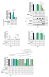
Reconstitution of necrosome interaction in S. cerevisiae. Maximal growth rates of yeast bearing expression plasmids encoding (A) single murine or human, or (B) human wild type necroptotic effector(s), were analyzed by monitoring changes in absorbance of yeast cultured in inducing and repressing liquid media. Expression of recombinant protein(s) were assessed by Western blot. Loading control: anti-hexokinase. (C) Yeast bearing expression plasmids encoding human necroptotic effector(s) were grown under inducing conditions for 24 h. Following galactose removal, plasma membrane integrity of each yeast culture was assessed by propidium iodide (PI) uptake. (D) The colony-forming abilities of post-induction cultures were expressed relative to the corresponding uninduced cultures. Cells were grown in inducing media for 24 h. Following galactose removal, the post-induction cultures were diluted and plated on solid repressing media and incubated for 2 days. Data present mean ± SEM of three independent assays in (AC) and five independent assays in (D). Differences in growth rates of (A) empty vector yeast versus those expressing each transgene, or (BD) sets of 2–3 transgenes versus individual transgenes or pairs of transgenes were compared using ANOVAs with Sidak corrections. *, p < 0.05; **, p < 0.01; ***, p < 0.001; ****, p < 0.0001; ns, not significant.
Figure 2
Figure 2
Reconstitution of necrosome interactions using RHIM-mutated RIPK3 in S. cerevisiae. Maximal growth rates of yeast-bearing expression plasmids encoding (A) human wild type and mutated RIPK3 proteins, or (B,E) human wild type MLKL, RIPK1 and/or RIPK3VQVG/AAAA, RIPK3VQVG/AAAA-D142N or RIPK3VQVG/AAAA-S227A, were analyzed by monitoring changes in absorbance of yeast cultured in inducing and repressing liquid media. (C) Yeast bearing expression plasmids encoding human wild-type MLKL and/or RIPK1, and/or RIPK3VQVG/AAAA were grown under inducing conditions for 24 h. Plasma membrane integrity of each yeast culture was assessed by propidium iodide (PI) uptake. (D) The clonogenic abilities of yeast transformants were evaluated by determining colony-forming ability of the culture relative before and after transgene induction for 24 h. Data present mean ± SEM of three (A,B,E) or four (C,D) independent assays. (A) Differences in growth rates of yeast expressing wild type RIPK3 and those expressing each mutated RIPK3 transgene were compared using ANOVAs with Sidak corrections. (BE) Statistical analysis of differences in growth rates, membrane integrity and clonogenic ability between transformants expressing sets of 2–3 transgenes versus individual transgenes or pairs of transgenes were calculated using ANOVAs with Sidak corrections. *, p < 0.05; **, p < 0.01; ***, p < 0.001; ns, not significant.
Figure 3
Figure 3
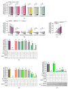
MLKL activation leads to its aggregation in yeast. (A) Subcellular distribution of C-terminal GFP-tagged MLKL in yeast after 24 h-induction in liquid media. Scale bars are 10 µm. Quantitative analyses of (B) yeast populations in terms of the subcellular distribution of GFP-tagged MLKL, and (C) subcellular localization of MLKLGFP puncta in yeast were assessed by confocal microscopy. Yeast expressing GFP or GFP-tagged MLKL with or without RIPK1 and/or RIPK3 were grown in inducing media for 24 h. At least 35 cells per yeast culture were counted in each independent assay. Graphs present mean ± SEM of three independent assays.
Figure 4
Figure 4
TC13172 and necrostatin inhibit necrosome interaction-induced yeast lethality. Maximal growth rates of yeast bearing (A) empty vectors or (B,C) expression plasmids encoding human wild-type MLKL, RIPK1 and RIPK3VQVG/AAAA in inducing versus repressing liquid media with different concentrations of indicated small molecule necroptotic inhibitors. Differences in growth rates of yeast in media either without drugs (-) or containing (A,B) 20, 40, 60, 80 or 100 µM of TC13172, NSA, GW806742X, necrostatin, GSK’872 or Dabrafenib; or (C) 4, 8, 12, 16 or 20 µM of TC13172 (increasing concentrations indicated by black triangle) were compared. Graphs present mean ± SEM of three independent assays. The effects of (D) 20 µM TC13172, (E) 100 µM necrostatin or (F) 100 µM GSK’872 on the growth rates of yeast expressing either MLKL, RIPK3, RIPK3VQVG/AAAA or RIPK1 or co-expression of two or three transgenes were assessed by monitoring the change in absorbance of yeast cultures in inducing and repressing media. The effect of GSK’872 on proliferation and MLKL phosphorylation of yeast bearing various transgenes were assayed (F). Differences in growth rates of untreated versus drug-treated yeast were compared using ANOVAs with Sidak corrections (AF). Graphs present mean ± SEM of three independent assays. *, p < 0.05; **, p < 0.01; ***, p < 0.001; ****, p < 0.0001; ns, not significant.
Figure 5
Figure 5
Viral necroptotic inhibitors reduce necrosome reconstitution-induced yeast lethality. (A,B) The effects of M45 on the growth of yeast in liquid media were analyzed by co-expressing necrosome effector(s) in yeast. Maximal growth rate of yeast bearing expression plasmids encoding human wild-type MLKL, RIPK1 and either RIPK3 (A,C), or RIPK3VQVG/AAAA (B,D), with and without M45 (A,B) or BAV_Rmil (C,D) in liquid media, were analyzed by monitoring the relative maximal changes in absorbance between yeast cultures in inducing and repressing media. Differences in growth rates of yeast expressing necrosome effectors with or without the viral inhibitors were compared using ANOVAs with Sidak corrections. Data present mean ± SEM of three independent assays. *, p < 0.05; **, p < 0.01; ***, p < 0.001; ****, p < 0.0001 ns, not significant.

Similar articles

Cited by

References

    1. Choi M.E., Price D.R., Ryter S., Choi A.M.K. Necroptosis: A crucial pathogenic mediator of human disease. JCI Insight. 2019;4:128834. doi: 10.1172/jci.insight.128834. - DOI - PMC - PubMed
    1. Dhuriya Y.K., Sharma D. Necroptosis: A regulated inflammatory mode of cell death. J. Neuroinflamm. 2018;15:1–9. doi: 10.1186/s12974-018-1235-0. - DOI - PMC - PubMed
    1. Kearney C.J., Martin S.J. An Inflammatory Perspective on Necroptosis. Mol. Cell. 2017;65:965–973. doi: 10.1016/j.molcel.2017.02.024. - DOI - PubMed
    1. Berghe T.V., Hassannia B., Vandenabeele P. An outline of necrosome triggers. Cell. Mol. Life Sci. 2016;73:2137–2152. doi: 10.1007/s00018-016-2189-y. - DOI - PMC - PubMed
    1. Cho Y.S., Challa S., Moquin D., Genga R., Ray T.D., Guildford M., Chan F.K.-M. Phosphorylation-Driven Assembly of the RIP1-RIP3 Complex Regulates Programmed Necrosis and Virus-Induced Inflammation. Cell. 2009;137:1112–1123. doi: 10.1016/j.cell.2009.05.037. - DOI - PMC - PubMed

LinkOut - more resources