. 2021 Jan 25;22(3):1172.
doi: 10.3390/ijms22031172.

Photodynamic and Cold Atmospheric Plasma Combination Therapy Using Polymeric Nanoparticles for the Synergistic Treatment of Cervical Cancer

Affiliations

Photodynamic and Cold Atmospheric Plasma Combination Therapy Using Polymeric Nanoparticles for the Synergistic Treatment of Cervical Cancer

Ji-Hui Ha et al. Int J Mol Sci. .

Abstract

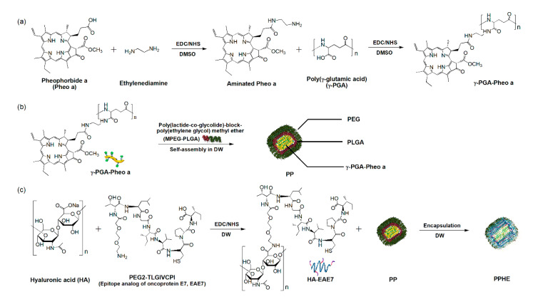
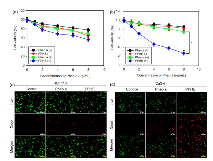
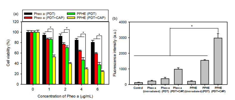
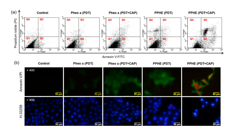
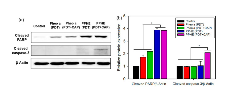
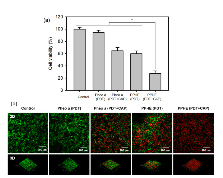
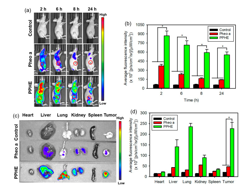
Integrating multi-modal therapies into one platform could show great promise in overcoming the drawbacks of conventional single-modal therapy and achieving improved therapeutic efficacy in cancer. In this study, we prepared pheophorbide a (Pheo a)/targeting ligand (epitope analog of oncoprotein E7, EAE7)-conjugated poly(γ-glutamic acid) (γ-PGA)/poly(lactide-co-glycolide)-block-poly(ethylene glycol) methyl ether (MPEG-PLGA)/hyaluronic acid (PPHE) polymeric nanoparticles via self-assembly and encapsulation method for the photodynamic therapy (PDT)/cold atmospheric plasma (CAP) combinatory treatment of human papillomavirus (HPV)-positive cervical cancer, thereby enhancing the therapeutic efficacy. The synthesized PPHE polymeric nanoparticles exhibited a quasi-spherical shape with an average diameter of 80.5 ± 17.6 nm in an aqueous solution. The results from the in vitro PDT efficacy assays demonstrated that PPHE has a superior PDT activity on CaSki cells due to the enhanced targeting ability. In addition, the PDT/CAP combinatory treatment more effectively inhibited the growth of cervical cancer cells by causing elevated intracellular reactive oxygen species generation and apoptotic cell death. Moreover, the three-dimensional cell culture model clearly confirmed the synergistic therapeutic efficacy of the PDT and the CAP combination therapy using PPHE on CaSki cells. Overall, these results indicate that the PDT/CAP combinatory treatment using PPHE is a highly effective new therapeutic modality for cervical cancer.

Keywords: cervical cancer; cold atmospheric plasma; combination therapy; photodynamic therapy; polymeric nanoparticles.


Conflict of interest statement

The authors declare no conflict of interest.

Figures

Figure 1
Figure 1
Preparation of the Pheo a/EAE7-conjugated γ-PGA/MPEG-PLGA/HA (PPHE) polymeric nanoparticles. Schematic diagram of the –synthetic process of the (a) Pheo a-conjugated γ-PGA (γ-PGA-Pheo a) prepared through the carbodiimide reaction to form amide linkage, (b) self-assembled γ-PGA-Pheo a/MPEG-PLGA (PP) nanoparticles, and (c) targeting ligand EAE7-decorated HA (HA-EAE7)-containing PPHE polymeric nanoparticles prepared via the amide bone formation and layer-by-layer (LBL) self-assembly method.
Figure 2
Figure 2
(a) Transmission electron microscopy (TEM) micrographs and (b) particle size distribution of the PPHE polymeric nanoparticles.
Figure 3
Figure 3
In vitro photodynamic therapy (PDT) efficacy of the free Pheo a and PPHE polymeric nanoparticles. Phototoxicity of different concentrations of the free Pheo a and PPHE polymeric nanoparticles on the (a) HCT116 and (b) CaSki cells before laser irradiation (Pheo a (−) and PPHE (−)) and after laser irradiation (Pheo a (+) and PPHE (+)) using a 671 nm laser (42 mW/cm2, 1 min) (n = 6). * p < 0.05 for comparison between two treatment groups. Fluorescence microscopy images of the (c) HCT116 and (d) CaSki cells after treatment with free Pheo a and PPHE polymeric nanoparticles (6 μg/mL Pheo a) and laser irradiation using a 671 nm laser (42 mW/cm2, 1 min). Live and dead cells were stained with calcein-AM (green) and EthD-1 (red), respectively.
Figure 4
Figure 4
(a) In vitro cytotoxicity of the PDT and cold atmospheric plasma (CAP) combination therapy on the CaSki cells evaluated by the MTT assay (n = 6). (b) DCF fluorescence intensity measured using a flow cytometer for determining the intracellular reactive oxygen species (ROS) level in the CaSki cells treated with free Pheo a and PPHE polymeric nanoparticles after laser irradiation and CAP treatment (n = 4). * p < 0.05 for comparison between two treatment groups.
Figure 5
Figure 5
(a) Cell death mechanism of the PDT and CAP combination therapy. The CaSki cells were treated with free Pheo a and PPHE polymeric nanoparticles (4 μg/mL Pheo a) for 18 h after irradiation with a 671 nm laser (42 mW/cm2, 1 min) and CAP (exposure time = 10 s). The cells were stained with annexin V and PI and analyzed on a flow cytometer. The upper-left (Q1), upper-right (Q2), lower-left (Q3), and lower-right (Q4) quadrants in each panel indicate the population of necrosis, late apoptosis, alive, and early apoptosis, respectively. (b) Morphology of the CaSki cells stained with annexin V/PI and H-33258 after treatment with free Pheo a and PPHE polymeric nanoparticles (4 μg/mL Pheo a), followed by irradiation using a 671 nm laser (42 mW/cm2, 1 min) and CAP (exposure time = 10 s).
Figure 6
Figure 6
(a) Representative western blots of the apoptosis-related proteins and (b) the quantified relative expression of the apoptosis-related proteins by densitometric analysis (n = 6). * p < 0.05 for comparison between two treatment groups. β-Actin was used as the loading control.
Figure 7
Figure 7
SEM micrographs of the surfaces and cross-sections of (a) the PLGA/γ-PGA/Pluronic 17R4 porous scaffolds with 3D bimodal pore structure fabricated via the thermally-induced phase separation (TIPS) method and (b) the CaSki cells grown on porous scaffolds for 15 days.
Figure 8
Figure 8
(a) In vitro cytotoxicity of the PDT and CAP combination therapy on the 3D cell culture model of the CaSki cells evaluated by the MTT assay (n = 4). * p < 0.05 for comparison between two treatment groups. (b) Fluorescence microscopy images of the 3D cell culture model of the CaSki cells after treatment with free Pheo a and PPHE polymeric nanoparticles (4 μg/mL Pheo a), followed by irradiation using a 671 nm laser (42 mW/cm2, 5 min) and CAP (exposure time = 50 s). Live and dead cells were stained with calcein-AM (green) and EthD-1 (red), respectively.
Figure 9
Figure 9
In vivo biodistribution of the free Ce6 and PPC nanoparticles. (a) In vivo time-dependent whole-body imaging of the tumor-bearing mice after the tail vein injection of free Pheo a and PPHE polymeric nanoparticles and (b) quantification of the average fluorescence signals in the tumor site. (c) Ex vivo fluorescence images of the tumor and normal organs (i.e., liver, lung, spleen, kidney, and heart) excised from the tumor-bearing mice at 24 h post-injection and (d) quantification of the average fluorescence signals of the tumor and normal organs. * p < 0.05 for comparison between two treatment groups.

Similar articles

Cited by

References

    1. Gao X., Jin Z., Tna X., Zhang C., Zou C., Zhang W., Ding J., Das B.C., Hitzeroth I.I., Debata P.R., et al. Hyperbranched poly(β-amino ester) based polyplex nanoparticles for delivery of CRISPR/Cas9 system and treatment of HPV infection associated cervical cancer. J. Control. Release. 2020;321:654–668. doi: 10.1016/j.jconrel.2020.02.045. - DOI - PubMed
    1. Alejo M., Alemany L., Clavero O., Quiros B., Vighi S., Seoud M., Cheng-Yang C., Garland S.M., Juanpere N., Lloreta J., et al. On behalf of RIS HPV TT study group. Contribution of human papillomavirus in neuroendocrine tumors from a series of 10,575 invasive cervical cancer cases. Papillomavirus Res. 2018;5:134–142. doi: 10.1016/j.pvr.2018.03.005. - DOI - PMC - PubMed
    1. Jin Y., Guan Z., Wang X., Wang Z., Zebg R., Xu L., Cao P. ALA-PDT promotes HPV-positive cervical cancer cells apoptosis and DCs maturation via miR-34a regulated HMGB1 exosomes secretion. Photodiagn. Photodyn. Ther. 2018;24:27–35. doi: 10.1016/j.pdpdt.2018.08.006. - DOI - PubMed
    1. Phelps W.C., Yee C.L., Munger K., Howley P.M. The human papillomavirus type 16 E7 gene encodes transactivation and transformation functions similar to those of adenovirus E1A. Cell. 1988;53:539–547. doi: 10.1016/0092-8674(88)90570-3. - DOI - PubMed
    1. Wieringa H.W., van der Zee A.G.J., de Vries E.G.E., van Vugt M.A.T.M. Breaking the DNA damage response to improve cervical cancer treatment. Cancer Treat. Rev. 2016;42:30–40. doi: 10.1016/j.ctrv.2015.11.008. - DOI - PubMed

MeSH terms