

Transcriptional Differences between Canine Cutaneous Epitheliotropic Lymphoma and Immune-Mediated Dermatoses
- PMID: 33504055
- PMCID: PMC7912288
- DOI: 10.3390/genes12020160
Transcriptional Differences between Canine Cutaneous Epitheliotropic Lymphoma and Immune-Mediated Dermatoses
Abstract
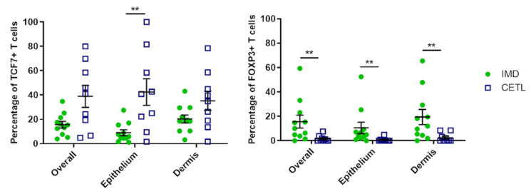
Canine cutaneous epitheliotropic T-cell lymphoma (CETL) and immune-mediated T-cell predominant dermatoses (IMD) share several clinical and histopathological features, but differ substantially in prognosis. The discrimination of ambiguous cases may be challenging, as diagnostic tests are limited and may prove equivocal. This study aimed to investigate transcriptional differences between CETL and IMD, as a basis for further research on discriminating diagnostic biomarkers. We performed 100bp single-end sequencing on RNA extracted from formalin-fixed and paraffin-embedded skin biopsies from dogs with CETL and IMD, respectively. DESeq2 was used for principal component analysis (PCA) and differential gene expression analysis. Genes with significantly different expression were analyzed for enriched pathways using two different tools. The expression of selected genes and their proteins was validated by RT-qPCR and immunohistochemistry. PCA demonstrated the distinct gene expression profiles of CETL and IMD. In total, 503 genes were upregulated, while 4986 were downregulated in CETL compared to IMD. RT-qPCR confirmed the sequencing results for 5/6 selected genes tested, while the protein expression detected by immunohistochemistry was not entirely consistent. Our study revealed transcriptional differences between canine CETL and IMD, with similarities to human cutaneous lymphoma. Differentially expressed genes are potential discriminatory markers, but require further validation on larger sample collections.
Keywords: Canis lupus familiaris; FFPE tissue; RNA sequencing; cutaneous T-cell lymphoma; cytotoxic dermatitis; dog; lupus erythematosus; skin disease; transcriptome.
Conflict of interest statement
The authors declare no conflict of interest. The funders had no role in study design, data collection and analysis, decision to publish, or preparation of the manuscript.
Figures


References
-
- Gross T.L., Ihrke P.J., Walder E.J., Affolter V.K., editors. Skin Disease of the Dog and Cat: Clinical and Histopathological Diagnosis. 2nd ed. Blackwell Publishing Ltd.; Ames, IA, USA: 2005.
-
- Miller W., Griffin C., Campbell K. Muller and Kirk’s Small Animal Dermatology. Saunders, Elsevier; St. Louis, MO, USA: 2013. Autoimmune and immune-mediated dermatoses; pp. 432–500.
Publication types
MeSH terms
LinkOut - more resources
Full Text Sources
Other Literature Sources
Medical
