Review
. 2021 Jan 25;10(2):90.
doi: 10.3390/biology10020090.

Anatomy and Neural Pathways Modulating Distinct Locomotor Behaviors in Drosophila Larva

Affiliations
Review

Anatomy and Neural Pathways Modulating Distinct Locomotor Behaviors in Drosophila Larva

Swetha B M Gowda et al. Biology (Basel). .

Abstract

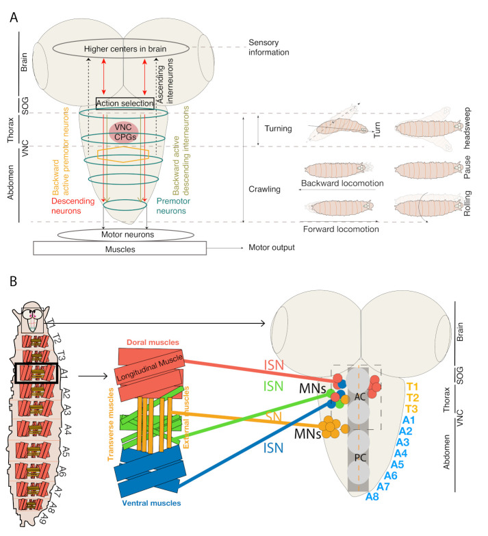
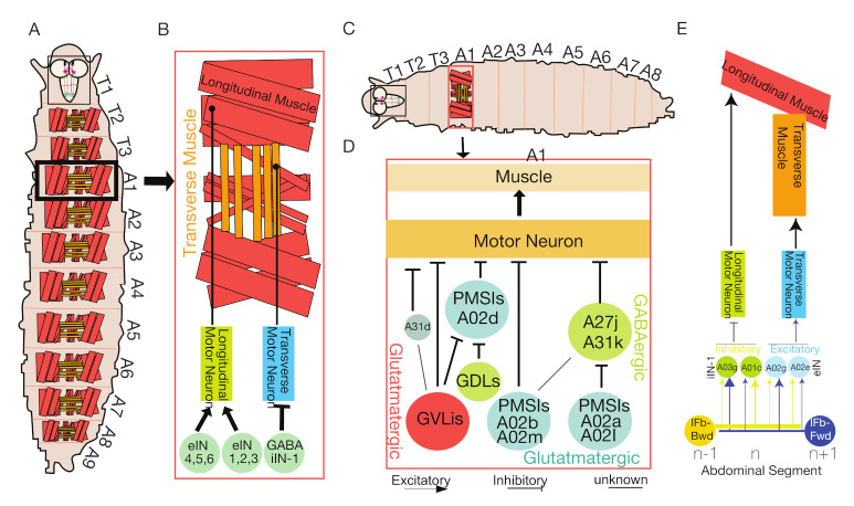
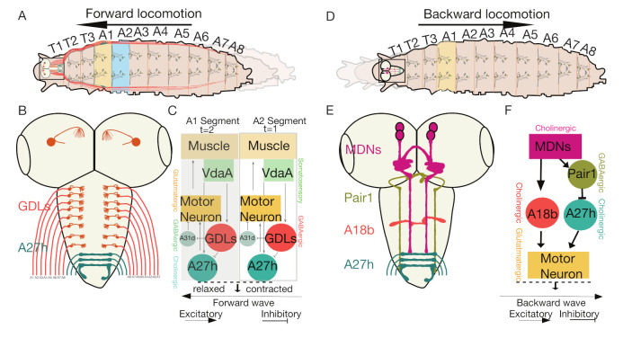
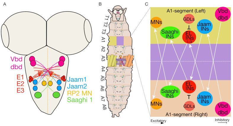
The control of movements is a fundamental feature shared by all animals. At the most basic level, simple movements are generated by coordinated neural activity and muscle contraction patterns that are controlled by the central nervous system. How behavioral responses to various sensory inputs are processed and integrated by the downstream neural network to produce flexible and adaptive behaviors remains an intense area of investigation in many laboratories. Due to recent advances in experimental techniques, many fundamental neural pathways underlying animal movements have now been elucidated. For example, while the role of motor neurons in locomotion has been studied in great detail, the roles of interneurons in animal movements in both basic and noxious environments have only recently been realized. However, the genetic and transmitter identities of many of these interneurons remains unclear. In this review, we provide an overview of the underlying circuitry and neural pathways required by Drosophila larvae to produce successful movements. By improving our understanding of locomotor circuitry in model systems such as Drosophila, we will have a better understanding of how neural circuits in organisms with different bodies and brains lead to distinct locomotion types at the organism level. The understanding of genetic and physiological components of these movements types also provides directions to understand movements in higher organisms.

Keywords: Drosophila larvae; brain circuits; information processing; locomotion; neural communication; sensory systems.


Conflict of interest statement

The authors declare no conflict of interest.

Figures

Figure 1
Figure 1
Schematics of topological organization, myotopic map and CNS regions involved in locomotion. (A) Schematics of organization of various types of neurons and thoracic and abdominal regions involved in various larval locomotor behaviors. (B) Schematic representation of Drosophila larval central nervous and neuromuscular system, which is divided into brain, subesophageal ganglia (SOG), and ventral nerve cord (VNC). In VNC, the positions of MNs cell bodies and representative muscle innervations are shown. MNs innervate abdominal hemisegments of each muscle through an intersegmental nerve (ISN, shown in red, blue and green), and segmental nerve (SN, shown in golden) which innervate internal and external (shown in golden) muscles, respectively. MN dendrite position in relation to their target muscles form a myotopic map. AC—Anterior commissure, PC—Posterior commissure (adapted and modified from [16]).
Figure 2
Figure 2
Interneuronal circuits regulating intrasegmental muscle contractions. (A) Schematic of Drosophila larva (not to scale) showing the anatomy of interneuronal circuits controlling coordinated longitudinal and transverse muscle segments. (B) Schematic showing neuromuscular connectivity (not to scale) of Longitudinal Muscles (LMs) (light red) and Transverse Muscles (TMs) (golden) that are innervated by longitudinal and transverse MNs, respectively (adapted and modified from [13,22]). (C) Schematic (not to scale) showing period positive median segmental interneurons (PMSIs) interneuronal circuits controlling MNs bursts duration. (D) PMSIs (A02b and A02m) provide inhibitory inputs to MNs. Other sets of PMSIs (A02a, A02I), presynaptic to GABAergic premotor neurons, remove inhibition from MNs after the relaxation of their target muscles. GVLIs inhibit MNs and control the termination of MNs bursting by being active at later phases of the locomotor cycle (adapted from [13,22]). (E) Intersegmental feedback neurons Ifb-Fwd and Ifb-Bwd neurons synapse to their target postsynaptic excitatory interneurons (A01c and A03g) that innervate transverse MNs and muscles and inhibitory interneurons (A02e, A02g and A14b) innervate longitudinal MNs and muscles for the regulation of bilateral motor coordination (adapted and modified from [22]).
Figure 3
Figure 3
Neuronal pathways controlling forward and backward locomotion. (A) Schematic of Drosophila larvae (top view) depicting forward locomotion. (B) Schematic showing the anatomy of the larval CNS depicting expression of GDL interneurons (red) and A27h neurons (green) in VNC. (C) Neural circuit in a hemisegment of muscles leading to the generation of forward locomotion (adapted and modified from [25]). (D) Schematic of Drosophila larvae (top view) depicting backward locomotion. (E) Schematic showing anatomy of larval CNS depicting expression of descending MDN, inhibitory Pair1, interneurons A18b, and premotor A27h neurons in brain and VNC of Drosophila larvae [23]. (F) A neural circuit in a hemisegment of muscle eliciting backward locomotion (adapted and modified from [23]).
Figure 4
Figure 4
Neural circuits are involved in bilateral motor coordination. (A) A schematic showing the anatomical location of cell bodies of neurons involved in bilateral motor coordination (adapted and modified from [50]). (B) A schematic showing a top view of larval body segments. The red/blue/green territories in the abdominal segment A1 represent muscle groups in each segment. Red represents dorsal LMs, blue represents TMs, and green represents ventral LMs. Sensory nerves in A2 abdominal segments and MNs output to muscles (colored diamond, circles, and squares). (C) The EL (Eve positive lateral) interneuron sends bilateral connections to Jaam interneurons, Saaghi interneurons and MNs (RP2-output neurons), receives inputs from proprioceptive sensory neurons (Vbd and dbd) and controls bilateral motor coordination. The ELs also receive inhibitory input from GABAergic GDL interneurons (adapted and modified from [82]).
Figure 5
Figure 5
Neural circuit involved in mechanosensory-nociception. (A) The neural circuitry involved in promoting hunching and bending in response to a gentle air puff (adapted and modified from [30]). (B) Schematic depicting larval hunching and bending in response to an air puff. (C) The neural circuitry involved in location-specific, gentle touch-mediated forward and backward locomotion (adapted and modified from [24]). (D) Schematic depicting touch-mediated forward and backward locomotion. (E) The neural circuitry involved in noxious, touch-mediated rolling behavior. (F) Schematic showing larval rolling in response to a noxious mechanical touch (>45 mN [104]) (adapted and modified from [24,29]).
Figure 6
Figure 6
The neural circuits involved in thermo-nociception-mediated bending and rolling. Neural circuits depicting multiple pathways that can be recruited when larvae experience noxious heat (adapted and modified from [31,105]).
Figure 7
Figure 7
Neural circuit involved in chemotaxis. (A) Schematic depicting larval turning towards attractive food odor. (B) Neural circuits showing chemotactic pathways involving either MB or LH leading to inhibition of forward locomotion and promoting turn towards odor (adapted and modified from [32,80]).
Figure 8
Figure 8
Schematics of neural circuit involved in phototaxis behavior. (A) Larval schematic showing head sweep behavior (B) Neural circuit showing phototaxis pathway, which mediate head sweep or turning behavior in response to low and intense light, respectively (adapted and modified from [125,127]).

Similar articles

Cited by

References

    1. Selverston A.I. Neural Control of Locomotion. Springer; Boston, MA, USA: 1976. Neuronal Mechanisms for Rhythmic Motor Pattern Generation in a Simple System; pp. 377–399. Advances in Behavioral Biology.
    1. Marder E., Calabrese R.L. Principles of rhythmic motor pattern generation. Physiol. Rev. 1996;76:687–717. doi: 10.1152/physrev.1996.76.3.687. - DOI - PubMed
    1. Grillner S. The motor infrastructure: From ion channels to neuronal networks. Nat. Rev. Neurosci. 2003;4:573–586. doi: 10.1038/nrn1137. - DOI - PubMed
    1. Yoon Y., Park J., Taniguchi A., Kohsaka H., Nakae K., Nonaka S., Ishii S., Nose A. System level analysis of motor-related neural activities in larval Drosophila. J. Neurogenet. 2019;33:179–189. doi: 10.1080/01677063.2019.1605365. - DOI - PubMed
    1. Jordan K.W., Carbone M.A., Yamamoto A., Morgan T.J., Mackay T.F.C. Quantitative genomics of locomotor behavior in Drosophila melanogaster. Genome Biol. 2007;8:R172. doi: 10.1186/gb-2007-8-8-r172. - DOI - PMC - PubMed

LinkOut - more resources