. 2021 Jan 25;22(3):1183.
doi: 10.3390/ijms22031183.

Solution Structure, Dynamics, and New Antifungal Aspects of the Cysteine-Rich Miniprotein PAFC

Affiliations

Solution Structure, Dynamics, and New Antifungal Aspects of the Cysteine-Rich Miniprotein PAFC

András Czajlik et al. Int J Mol Sci. .

Abstract

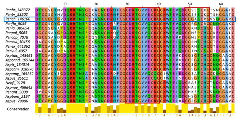
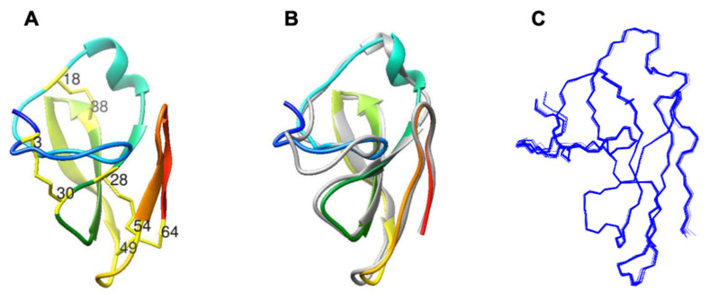
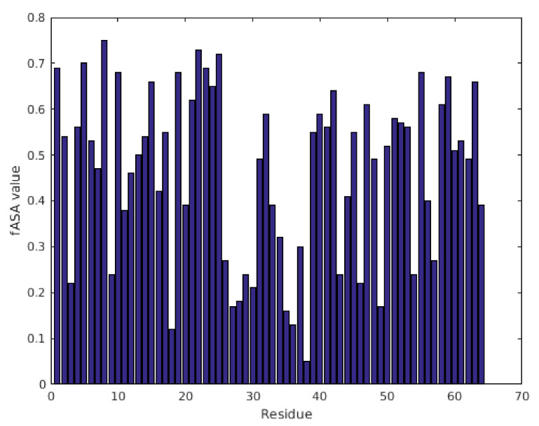
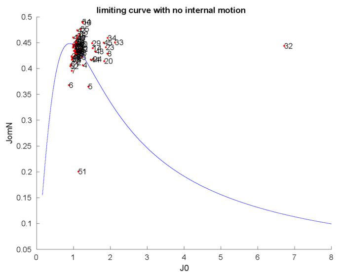
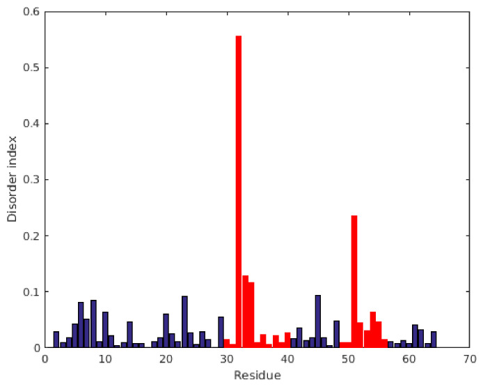
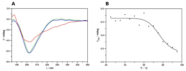
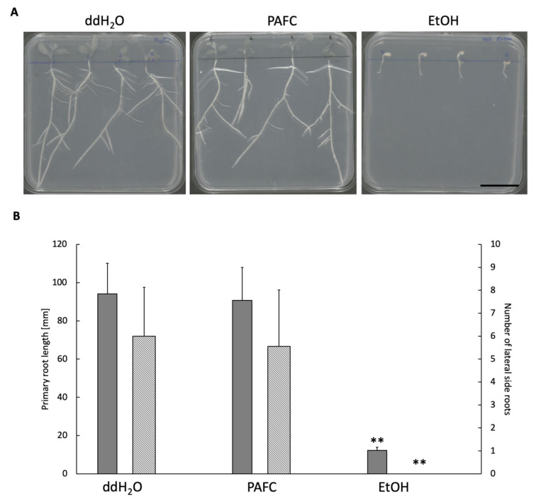
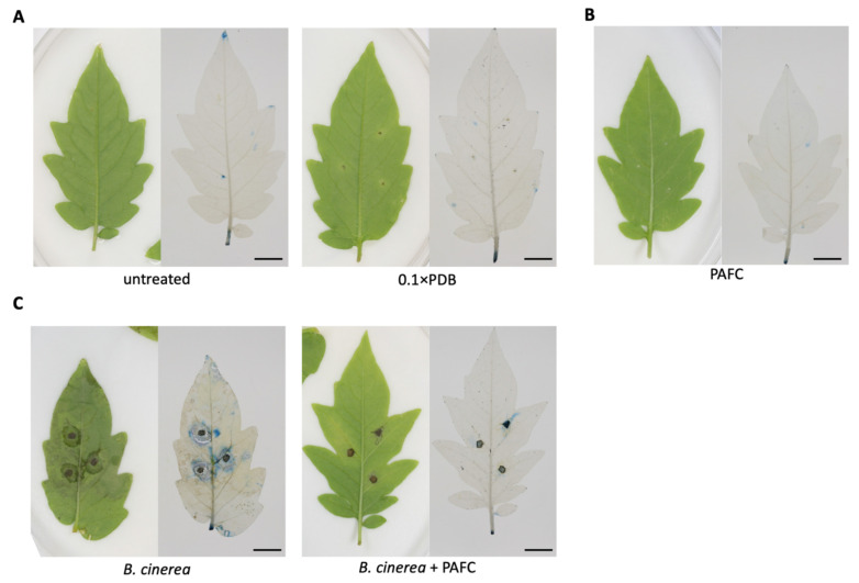
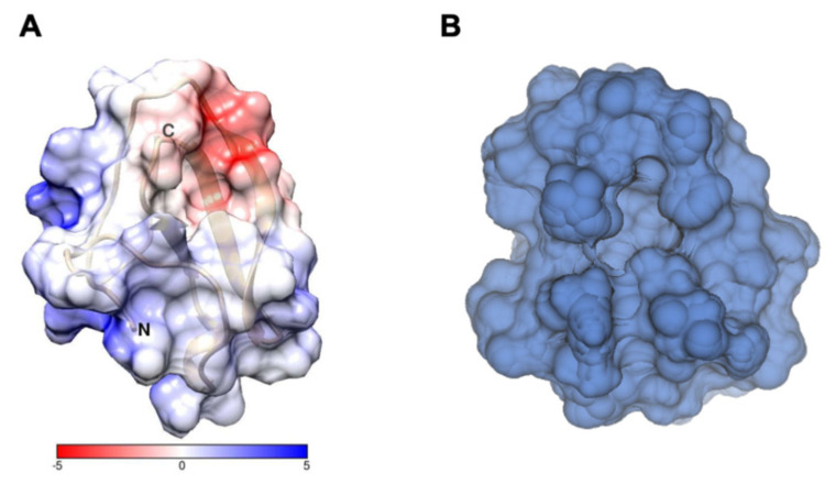
The genome of Penicillium chrysogenum Q176 contains a gene coding for the 88-amino-acid (aa)-long glycine- and cysteine-rich P. chrysogenum antifungal protein C (PAFC). After maturation, the secreted antifungal miniprotein (MP) comprises 64 aa and shares 80% aa identity with the bubble protein (BP) from Penicillium brevicompactum, which has a published X-ray structure. Our team expressed isotope (15N, 13C)-labeled, recombinant PAFC in high yields, which allowed us to determine the solution structure and molecular dynamics by nuclear magnetic resonance (NMR) experiments. The primary structure of PAFC is dominated by 14 glycines, and therefore, whether the four disulfide bonds can stabilize the fold is challenging. Indeed, unlike the few published solution structures of other antifungal MPs from filamentous ascomycetes, the NMR data indicate that PAFC has shorter secondary structure elements and lacks the typical β-barrel structure, though it has a positively charged cavity and a hydrophobic core around the disulfide bonds. Some parts within the two putative γ-core motifs exhibited enhanced dynamics according to a new disorder index presentation of 15N-NMR relaxation data. Furthermore, we also provided a more detailed insight into the antifungal spectrum of PAFC, with specific emphasis on fungal plant pathogens. Our results suggest that PAFC could be an effective candidate for the development of new antifungal strategies in agriculture.

Keywords: Penicillium chrysogenum; antifungal protein PAFC; dynamics; nuclear magnetic resonance; plant protection; solution structure; γ-core motif.


Conflict of interest statement

The authors declare no conflicts of interest.

Figures

Figure 1
Figure 1
ClustalW multiple alignment of the mature, P. brevicompactum “bubble protein” BP-clade-specific MPs of Eurotiomycetes [21]. P. chrysogenum PAFC (Pench_146100) is framed in blue. The two conserved γ-core motifs found in the members of this clade are framed in red. The abbreviations of the full species names and the protein accession number are indicated: Penbr (P. brevicompactum), Pench (P. chrysogenum), Penfla (Penicillium falvigenum) Penla (Penicillium lanosocoeruleum), Penpol (Penicillium polonicum), Pencop (Penicillium coprophilum), Penswi (Penicillium swiecickii), Penex (Penicillium expansum), Penvul (Penicillium vulpinum), Asptaic (Aspergillus taichungensis), Aspcand (Aspergillus candidus), Asptr (Aspergillus triticus), Aspcam (Aspergillus campestris), Aspeamy (Aspergillus amylovorus), Aspve (Aspergillus versicolor), Neofi (Neosartorya fischeri), Aspnov (Aspergillus novofumigatus), Penant (Penicillium antarticum), and Aspbom (Aspergillus bombycis). The color coding of aa was applied according to ClustalX. The conservation between the respective sequences based on the ClustalW2 Multiple Sequence Alignment tool [26] is indicated at the bottom.
Figure 2
Figure 2
Comparison of the structures of Penicillium spp. MPs. (A) The NMR solution structure of the PAFC (pdb code 6TRM). The position of the Cys residues is indicated, and the disulfide bonds are labeled as yellow sticks. (B) Superimposed structures of the PAFC (pdb code 6TRM) and the P. brevicompactum BP (gray color; pdb code: 1UOY) using Chimera visualization software [33]. (C) Backbone NMR conformational ensemble of 20 structures of PAFC.
Figure 3
Figure 3
fASA values of PAFC calculated from 13C chemical shift data show residue-by-residue hydrophobicity of the structure. Residues with fASA values below 0.25 are considered strongly buried, while fASA values above 0.75 indicate exposure to solvent.
Figure 4
Figure 4
Reduced spectral density mapping of NH mobilities in PAFC (298 K). The limiting continuous curve represents the absence of internal motion, as calculated by τc = 3.14 ns global correlation time. Spectral densities (the strength of fluctuating radiofrequency fields from molecular rotational diffusion) at 15N frequency shown as a function of such fields close to zero frequency (slow-motion regime) [40]. JomN on the vertical axis shows the spectral densities at 15N frequency, while J0 on the horizontal axis is proportional to spectral densities close to zero frequency. The units of the two axes should be understood as 10−9 (s rad−1).
Figure 5
Figure 5
Disorder index (DI), as obtained directly from reduced spectral density mapping of 15N NMR relaxation data. The DI is just derived from Figure 4 by calculating the geometrical (shortest) distances of the points from the solid (limiting) curve. The γ-core regions are labeled as red bars, while other residues are blue.
Figure 6
Figure 6
ECD spectra and thermal unfolding curve of PAFC. (A) Spectra in the 185–260 nm region were measured at 25 °C (green), 95 °C (red), and again 25 °C (blue) after refolding. (B) Thermal unfolding of PAFC, followed by ECD spectra at 229 nm. Tm = 77 °C.
Figure 7
Figure 7
The primary structure of PAFC and the derived synthetic γ-core peptides. The aa sequence of PAFC is indicated by a one-letter code. The predicted levomeric γ-core motifs in the center and the C-terminus are highlighted in red. Below, sequences of the N-terminally acetylated (Ac-) synthetic peptides PCγ15, PCγ17, and PCγC-terminal are indicated.
Figure 8
Figure 8
Vegetative growth and root development of Medicago truncatula A-17. (A) Morphology of plant seedlings and (B) primary root length (gray bars) and number of lateral roots (hatched bars) after daily treatment with 1 mg mL−1 of PAFC for 10 days at 25 °C under continuous illumination (1200 lux) compared to the ddH2O- and 70% (v/v) ethanol-treated controls, respectively. Scale bar, 30 mm. Significant difference in (B) was evaluated with the two-sample t-test and is indicated with ** (p < 0.005).
Figure 9
Figure 9
PAFC tolerance of tomato plant leaves and protective effect of PAFC against B. cinerea infection. The phenotype of leaves is shown in the left panels and damage evaluation with Evan’s blue staining in the right panels. (A) Leaves were either left untreated or treated with 10 µL of 0.1 × PDB (negative controls). (B) For toxicity testing, leaves were treated with 10 µL of PAFC (1 mg mL−1). (C) Plant protection efficacy was evaluated with leaves infected with 10 µL of B. cinerea conidia (107 conidia mL−1) (infection control) and conidia mixed with PAFC (1 mg mL−1). Plants were then further incubated for four days at 23 °C under photoperiodic day/night simulation (12/12 h with or without illumination at 1200 lux) and then harvested for staining. Scale bar, 10 mm.
Figure 10
Figure 10
(A) Electrostatic potential surface of PAFC, as calculated [48] from the 6TRM structure deposited in pdb. Red means negative, and blue means positive surfaces. The scale is in kJ/mol/e. (B) Front view of cavity B of PAFC, formed predominantly by residues 3, 12, 13, 14, 28, 29, 30, 31, 33, 34, 35, 36, 44, 45, and 48.

Similar articles

Cited by

References

    1. Fisher M.C., Gurr S.J., Cuomo C.A., Blehert D.S., Jin H.L., Stukenbrock E.H., Stajich J.E., Kahmann R., Boone C., Denning D.W., et al. Threats posed by the fungal kingdom to humans, wildlife, and agriculture. mBio. 2020;11 doi: 10.1128/mBio.00449-20. - DOI - PMC - PubMed
    1. Hancock R.E.W., Sahl H.-G. Antimicrobial and host-defense peptides as new anti-infective therapeutic strategies. Nat. Biotechnol. 2006;24:1551–1557. doi: 10.1038/nbt1267. - DOI - PubMed
    1. Brown K.L., Hancock R.E.W. Cationic host defense (antimicrobial) peptides. Curr. Opin. Immunol. 2006;18:24–30. doi: 10.1016/j.coi.2005.11.004. - DOI - PubMed
    1. Zasloff M. Antimicrobial peptides of multicellular organisms. Nature. 2002;415:389–395. doi: 10.1038/415389a. - DOI - PubMed
    1. Van der Weerden N.L., Bleackley M.R., Anderson M.A. Properties and mechanisms of action of naturally occurring antifungal peptides. Cell. Mol. Life Sci. 2013;70:3545–3570. doi: 10.1007/s00018-013-1260-1. - DOI - PMC - PubMed

Supplementary concepts

LinkOut - more resources