. 2021 Jan 25;26(3):625.
doi: 10.3390/molecules26030625.

Coupling of a Novel TIMP3 Peptide to Carboxypeptidase G2 for Pro-Drug Activation at the Tumour Site

Affiliations

Coupling of a Novel TIMP3 Peptide to Carboxypeptidase G2 for Pro-Drug Activation at the Tumour Site

Mohammed S Aldughaim et al. Molecules. .

Abstract

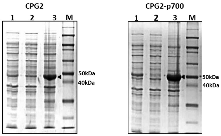
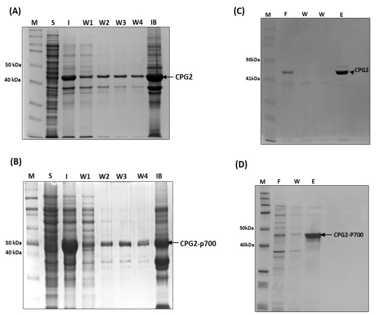
Broad-spectrum cytotoxic drugs have been used in cancer therapy for decades. However, their lack of specificity to cancer cells often results in serious side-effects, limiting efficacy. For this reason, antibodies have been used to attempt to specifically target cytotoxic drugs to tumours. One such approach is antibody-directed enzyme prodrug therapy (ADEPT) which uses a tumour-directed monoclonal antibody, coupled to an enzyme, to convert a systemically administered non-toxic prodrug into a toxic one only at the tumour site. Among the main drawbacks of ADEPT is the immunogenicity of the antibody-enzyme complex, which is exacerbated by slow clearance due to size, hence limiting repeated administration. Additionally, the mono-specificity of the antibody could potentially result in drug resistance with repeated administration. We have identified a novel short peptide sequence, p700, derived from a human tissue inhibitor of metalloproteinases-3 (TIMP-3), which binds to and inhibits a number of tyrosine kinase growth factor receptors (VEGFRs1-3, FGFRs 1-4 and PDGFRα) which are known to be upregulated in many tumours and tumour vasculature. In this report, we fused p700 to His-tagged, codon-optimised, carboxypeptidase G2 (CPG2). CPG2 is a bacterial enzyme used in ADEPT, which activates potent nitrogen-mustard pro-drugs by removal of an inhibitory glutamic acid residue. Recombinant CPG2-p700 was highly expressed in Escherichia coli and successfully purified by nickel affinity chromatography. Biolayer interferometry showed that CPG2-p700 had a 100-fold increase in binding affinity for VEGFR2 compared with CPG2 alone and retained its catalytic activity, as determined by methotrexate cleavage. In the presence of CPG2-p700, the ZD2676P pro-drug showed significant cytotoxicity for 4T1 cells compared with prodrug alone or CPG2 alone. p700 is, therefore, a potentially useful alternative to monoclonal antibodies for enzyme pro-drug therapy and could equally be used for effective delivery of other cytotoxic drugs to tumour tissue.

Keywords: TIMP3; VEGFR2; angiogenesis; carboxypeptidase G2.


Conflict of interest statement

The authors declare no conflict of interest.

Figures

Figure 1
Figure 1
Solubility test to determine localisation of recombinant CPG2 protein expression in BL21 E. coli. Coomassie Blue staining following SDS-PAGE of samples of whole cell lysate (lane 1), supernatant (lane 2) and re-suspended pellet (lane 3) of IPTG induced BL21 E. coli expressing CPG2 and CPG2-p700 proteins, respectively, from left to right, along with protein standards (ladder). The arrowheads indicate over-expressed CPG2 and CPG2-p700 proteins at the expected sizes (45 and 48 kDa, respectively) in the re-suspended pellets.
Figure 2
Figure 2
Large-scale expression of CPG2 recombinant proteins in BL21 E. coli. Panel (A) Coomassie blue staining following SDS-PAGE of paired samples (whole cell lysate) taken pre- (u) and 3 h post-IPTG induction (3 h) of CPG2, and CPG2-p700 recombinant protein expression, respectively, from left to right, compared with a ladder (extreme left). The arrowheads highlight the protein bands of the expected sizes (45 and 48 kDa). Panel (B) Western blotting analysis using antihistidine antibody on identical samples corresponding to the top panel, compared with a protein ladder (extreme left). Histidine-tagged recombinant proteins of the expected sizes (45 and 48 kDa) were detected in post-induction samples, but not in corresponding pre-induction samples.
Figure 3
Figure 3
Purification of recombinant CPG2 proteins on nickel-chelate resin from urea washes derived from the insoluble fraction of BL21 E. coli cell lysates. (A) CPG2 and (B) CPG2-p700 coomassie blue stained SDS-PAGE gels. Lanes are of standard protein ladder; soluble fraction; insoluble fraction; four sequential urea washes; and remaining inclusion body, respectively, of IPTG-induced BL21 E. coli cells expressing. Pooled washes (1 to 4) from the insoluble fraction of lysates derived the IPTG-induced BL21 E. coli cells expressing His-tagged recombinant (C) CPG2 and (D) CPG2-p700 (right panel) were purified using a Ni2+ chelate column. Lanes (left to right) represent Coomassie Blue staining following SDS-PAGE of standard protein ladder, initial flow-through, wash and eluent (purified recombinant protein).
Figure 4
Figure 4
Confirmation of the zinc-dependent enzymatic activity of purified recombinant CPG2 proteins. Recombinant proteins were incubated with methotrexate at 37 °C for 0–60 min. The absorbance of each reaction at 320 nm was plotted against time. A similar reduction in absorbance (representing catalysis of MTX) was seen for both CPG2 (A) and CPG2-p700 (B) in the presence of ZnSO4 but not in its absence nor when the zinc chelating agent, EDTA, was added.
Figure 5
Figure 5
A representative biolayer interferometry experiment comparing the interaction between recombinant CPG2 or CPG2-p700 and VEGFR2. The binding signal is the wavelength shift detected by the BLItz machine and corresponds to the change in thickness of the biolayer as a result of the interaction between the recombinant proteins (CPG2 or CPG2-p700) and a VEGFR2-coated biosensor and is plotted against the duration of the interactions. Four runs are shown with PBS only loaded (green line), VEGFR2 only loaded (purple line) and VEGFR2 loaded followed by CPG2 (yellow line) or CPG2-p700 (green line). The first loading curve is the binding of VEGFR2-Fc to the protein A coated sensor. The sensor was then washed by dipping into PBS (baseline) before dipping into the CPG2 protein solutions (association). The sensor was then transferred to PBS and dissociation measured. Binding affinity (dissociation constant, Kd) was calculated using the BLItz software based on five repeats using increasing concentrations (300 nM to 10 mM) of recombinant proteins. CPG2-p700 showed a 100-fold increase in binding affinity compared with CPG2.
Figure 6
Figure 6
The effect of increasing concentrations of prodrug ZD2676P on the viability of the mouse breast cancer cell line 4T1 in the presence or absence of CPG2 or CPG2-p700. Data are means ± SEM; n = 3 (** = p < 0.01, *** = p < 0.001) indicates significance, two-way ANOVA, multiple comparison test.

References

    1. Chen Y., Jia Y., Song W., Zhang L. Therapeutic potential of nitrogen mustard based hybrid molecules. Front. Pharmacol. 2018;9:1453. doi: 10.3389/fphar.2018.01453. - DOI - PMC - PubMed
    1. Sharma S.K., Bagshawe K.D. Antibody directed enzyme prodrug therapy (ADEPT): Trials and tribulations. Adv. Drug Deliv. Rev. 2017;118:2–7. doi: 10.1016/j.addr.2017.09.009. - DOI - PubMed
    1. Sharma S.K., Bagshawe K.D. Translating antibody directed enzyme prodrug therapy (ADEPT) and prospects for com-bination. Expert Opin. Biol. Ther. 2017;17:1–13. doi: 10.1080/14712598.2017.1247802. - DOI - PubMed
    1. Sharma S.K., Bagshawe K.D., Melton R.G., Sherwood R.F. Human immune response to monoclonal antibody-enzyme conjugates in ADEPT pilot clinical trial. Cell Biophys. 1992;21:109–120. doi: 10.1007/BF02789482. - DOI - PubMed
    1. Goda S.K., Rashidi F.A.B., Fakharo A.A., Al-Obaidli A. Functional overexpression and purification of a codon optimized synthetic glucarpidase (carboxypeptidase G2) in Escherichia coli. Protein J. 2009;28:435–442. doi: 10.1007/s10930-009-9211-2. - DOI - PubMed

MeSH terms

LinkOut - more resources