

Retinal Vascular Endothelial Cell Dysfunction and Neuroretinal Degeneration in Diabetic Patients
- PMID: 33504108
- PMCID: PMC7866162
- DOI: 10.3390/jcm10030458
Retinal Vascular Endothelial Cell Dysfunction and Neuroretinal Degeneration in Diabetic Patients
Abstract
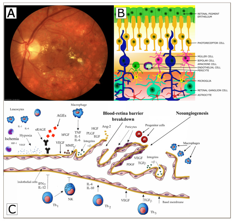
Diabetes mellitus (DM) has become a vital societal problem as epidemiological studies demonstrate the increasing incidence of type 1 and type 2 diabetes. Lesions observed in the retina in the course of diabetes, referred to as diabetic retinopathy (DR), are caused by vascular abnormalities and are ischemic in nature. Vascular lesions in diabetes pertain to small vessels (microangiopathy) and involve precapillary arterioles, capillaries and small veins. Pericyte loss, thickening of the basement membrane, and damage and proliferation of endothelial cells are observed. Endothelial cells (monolayer squamous epithelium) form the smooth internal vascular lining indispensable for normal blood flow. Breaking its continuity initiates blood coagulation at that site. The endothelium controls the process of exchange of chemical substances (nutritional, regulatory, waste products) between blood and the retina, and blood cell passing through the vascular wall. Endothelial cells produce biologically active substances involved in blood coagulation, regulating vascular wall tension and stimulating neoangiogenesis. On the other hand, recent studies have demonstrated that diabetic retinopathy may be not only a microvascular disease, but is a result of neuroretinal degeneration. Neuroretinal degeneration appears structurally, as neural apoptosis of amacrine and Muller cells, reactive gliosis, ganglion cell layer/inner plexiform (GCL) thickness, retinal thickness, and retinal nerve fiber layer thickness, and a reduction of the neuroretinal rim in minimum rim width (MRW) and functionally as an abnormal electroretinogram (ERG), dark adaptation, contrast sensitivity, color vision, and microperimetric test. The findings in early stages of diabetic retinopathy may precede microvascular changes of this disease. Furthermore, the article's objective is to characterize the factors and mechanisms conducive to microvascular changes and neuroretinal apoptosis in diabetic retinopathy. Only when all the measures preventing vascular dysfunction are determined will the risk of complications in the course of diabetes be minimized.
Keywords: diabetes; diabetic retinopathy; dysfunction; endothelium; retinal neurodegeneration; retinal vessels.
Conflict of interest statement
The authors declare no conflict of interest.
Figures
References
-
- Hartwig S., Greiser K.H., Medenwald D., Tiller D., Herzog B., Schipf S., Ittermann T., Völzke H., Müller G., Haerting J., et al. Association of change of anthropometric measurements with incident type 2 diabetes mellitus: A pooled analysis of the prospective population-based CARLA and SHIP Cohort Studies. Medicine. 2015;94:e1394. doi: 10.1097/MD.0000000000001394. - DOI - PMC - PubMed
-
- Islam M.S. Diabetes: From Research to Clinical Practice. Adv. Exp. Med. Biol. 2020;1307:1–5. - PubMed
-
- Imperatore G., Boyle J.P., Thompson T.J., Case D., Dabelea D., Hamman R.F., Lawrence J.M., Liese A.D., Liu L.L., Mayer-Davis E.J., et al. Projections of type 1 and type 2 diabetes burden in the U.S. population aged <20 years through 2050: Dynamic modeling of incidence, mortality, and population growth. Diabetes Care. 2012;35:2515–2520. doi: 10.2337/dc12-0669. - DOI - PMC - PubMed
-
- World Health Organization . Prevention of Blindness from Diabetes Mellitus: Report of WHO Consultation in Geneva, Switzerland, 9–11 November 2005. World Health Organization; Geneva, Switzerland: 2006.
Publication types
LinkOut - more resources
Full Text Sources
Other Literature Sources
