

Syncytins expressed in human placental trophoblast
- PMID: 33504453
- PMCID: PMC8280254
- DOI: 10.1016/j.placenta.2021.01.006
Syncytins expressed in human placental trophoblast
Abstract
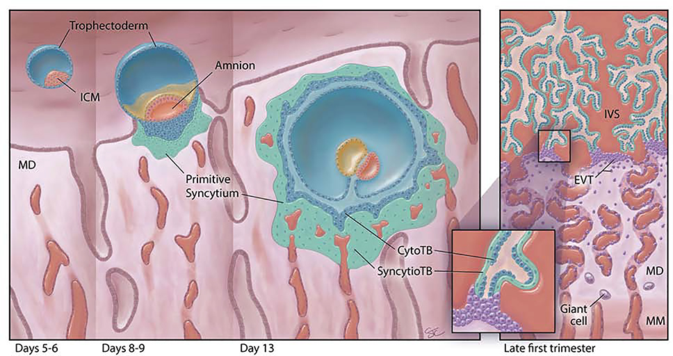
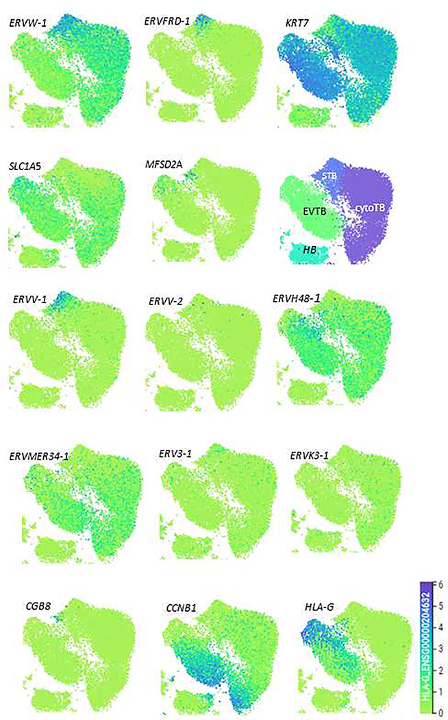
Three versions of syncytiotrophoblast exist in the human placenta: an invasive type associated with the implanting conceptus, non-invasive villous type of definitive placenta, and placental bed giant cells. Syncytins are encoded by modified env genes of endogenous retroviruses (ERV), but how they contribute functionally to placental syncytial structures is unclear. A minimum of eight genes (ERVW1, ERVFRD-1, ERVV-1, ERVV-2, ERVH48-1, ERVMER34-1, ERV3-1, & ERVK13-1) encoding syncytin family members are expressed in human trophoblast, the majority from implantation to term. ERVW1 (Syncytin 1) and ERVFRD-1 (Syncytin 2) are considered the major fusogens, but, when the expression of their genes is analyzed by single cell RNAseq in first trimester placenta, their transcripts are distinctly patterned and also differ from those of their proposed binding partners, SLC1A5 and MFSD2A, respectively. ERVRH48-1 (suppressyn or SUPYN) and ERVMER34-1 are probable negative regulators of fusion and co-expressed, primarily in cytotrophoblast. The remaining genes and their products have been little studied. Syncytin expression is a feature of placental development in almost all eutherian mammals studied, in at least one marsupial, and in viviparous lizards, which lack the trophoblast lineage. Their expression has been inferred to be essential for pregnancy success in the mouse. All the main human ERV genes arose following independent retroviral insertion events, none of which trace back to the divergence of eutherians and metatherians (marsupials). While syncytins may be crucial for placental development, it seems unlikely that they helped orchestrate the divergence of eutherians and marsupials.
Keywords: Endogenous retrovirus; Extravillous trophoblast; Fusogen; Placental evolution; Suppressyn; Syncytiotrophoblast.
Copyright © 2021 Elsevier Ltd. All rights reserved.
Conflict of interest statement
Declarations of interest: none
The authors declare no conflicts of interest.
Figures
Comment in
-
Comment to "Syncytin expression in human placental trophoblast".Placenta. 2022 Aug;126:26. doi: 10.1016/j.placenta.2022.05.010. Epub 2022 May 21. Placenta. 2022. PMID: 35691202 No abstract available.
References
-
- Vento-Tormo R, Efremova M, Botting RA, Turco MY, Vento-Tormo M, Meyer KB, Park JE, Stephenson E, Polanski K, Goncalves A, Gardner L, Holmqvist S, Henriksson J, Zou A, Sharkey AM, Millar B, Innes B, Wood L, Wilbrey-Clark A, Payne RP, Ivarsson MA, Lisgo S, Filby A, Rowitch DH, Bulmer JN, Wright GJ, Stubbington MJT, Haniffa M, Moffett A, Teichmann SA, Single-cell reconstruction of the early maternal-fetal interface in humans, Nature 563(7731) (2018) 347–353. - PMC - PubMed
-
- Hernandez JM, Podbilewicz B, The hallmarks of cell-cell fusion, Development 144(24) (2017) 4481–4495. - PubMed
-
- Hertig AT, The primary human oocyte: some observations on the fine structure of Balbiani’s vitelline body and the origin of the annulate lamellae, Am J Anat 122(1) (1968) 107–37. - PubMed
Publication types
MeSH terms
Substances
Grants and funding
LinkOut - more resources
Full Text Sources
Other Literature Sources
