

Comparative cranial biomechanics in two lizard species: impact of variation in cranial design
- PMID: 33504585
- PMCID: PMC7970069
- DOI: 10.1242/jeb.234831
Comparative cranial biomechanics in two lizard species: impact of variation in cranial design
Abstract
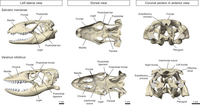
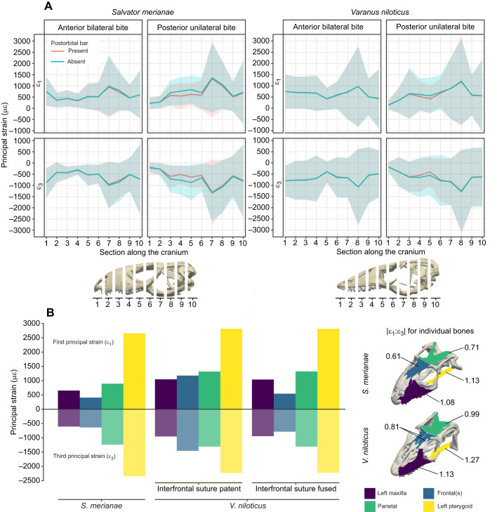
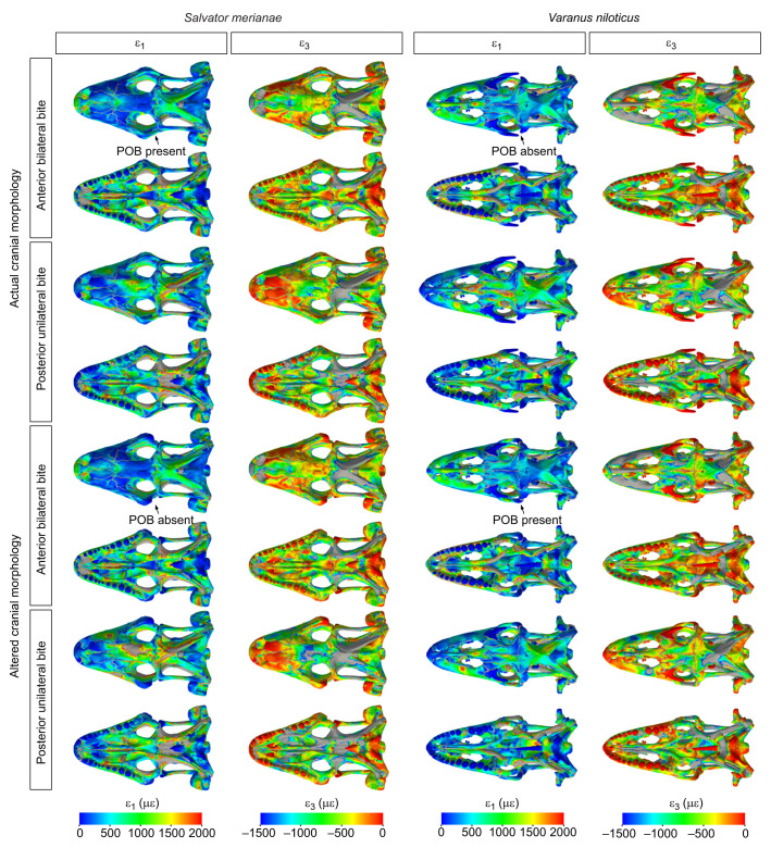
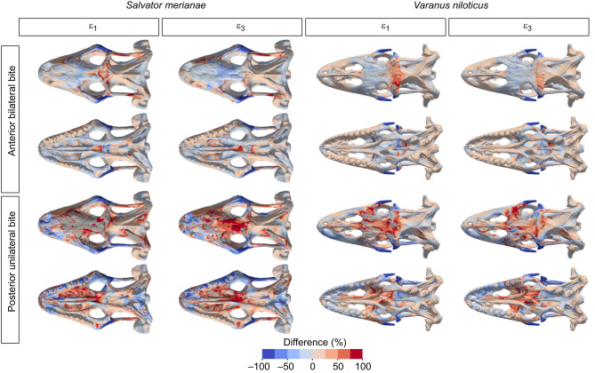
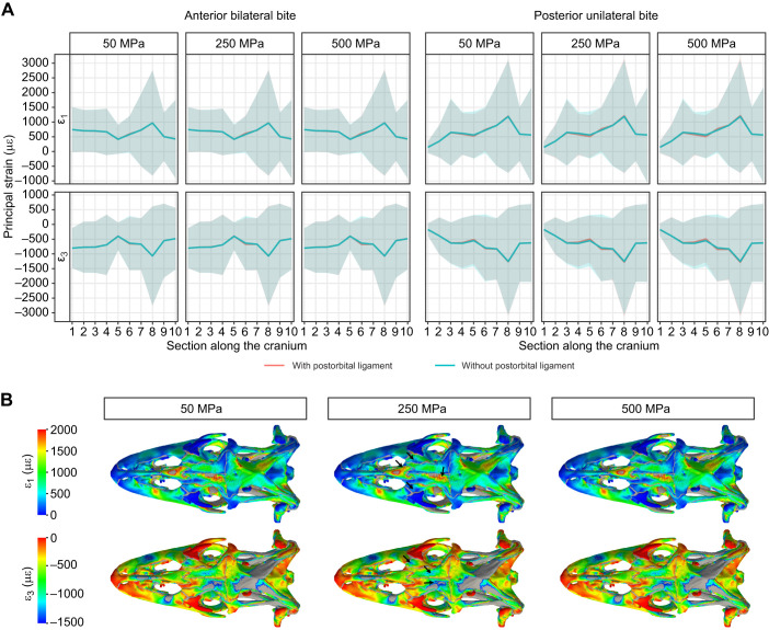
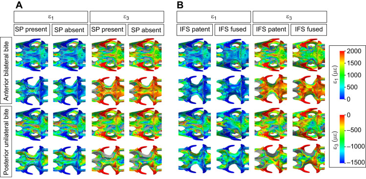
Cranial morphology in lepidosaurs is highly disparate and characterised by the frequent loss or reduction of bony elements. In varanids and geckos, the loss of the postorbital bar is associated with changes in skull shape, but the mechanical principles underlying this variation remain poorly understood. Here, we sought to determine how the overall cranial architecture and the presence of the postorbital bar relate to the loading and deformation of the cranial bones during biting in lepidosaurs. Using computer-based simulation techniques, we compared cranial biomechanics in the varanid Varanus niloticus and the teiid Salvator merianae, two large, active foragers. The overall strain magnitude and distribution across the cranium were similar in the two species, despite lower strain gradients in V. niloticus In S. merianae, the postorbital bar is important for resistance of the cranium to feeding loads. The postorbital ligament, which in varanids partially replaces the postorbital bar, does not affect bone strain. Our results suggest that the reduction of the postorbital bar impaired neither biting performance nor the structural resistance of the cranium to feeding loads in V. niloticus Differences in bone strain between the two species might reflect demands imposed by feeding and non-feeding functions on cranial shape. Beyond variation in cranial bone strain related to species-specific morphological differences, our results reveal that similar mechanical behaviour is shared by lizards with distinct cranial shapes. Contrary to the situation in mammals, the morphology of the circumorbital region, calvaria and palate appears to be important for withstanding high feeding loads in these lizards.
Keywords: Feeding; Finite element analysis; Lepidosauria; Multibody dynamic analysis; Skull; Squamata.
© 2021. Published by The Company of Biologists Ltd.
Conflict of interest statement
Competing interestsThe authors declare no competing or financial interests.
Figures
References
-
- Ahrens, J., Geveci, B. and Law, C. (2005). ParaView: an end-user tool for large-data visualization. In Visualization Handbook (ed. Johnson C. R.), pp. 717-731. Burlington, USA: Butterworth-Heinemann.
-
- Burbrink, F. T., Grazziotin, F. G., Pyron, R. A., Cundall, D., Donnellan, S., Irish, F., Keogh, J. S., Kraus, F., Murphy, R. W., Noonan, B.et al. (2020). Interrogating genomic-scale data for Squamata (lizards, snakes, and amphisbaenians) shows no support for key traditional morphological relationships. Syst. Biol. 69, 502-520. 10.1093/sysbio/syz062 - DOI - PubMed
-
- Cartmill, M. (1980). Morphology, function, and evolution of the anthropoid postorbital septum. In Evolutionary Biology of the New World Monkeys and Continental Drift (ed. Ciochon R. L. and Chiarelli A. B.), pp. 243-274. Boston, USA: Springer US.
Publication types
MeSH terms
Grants and funding
- BB/M010287/1/BB_/Biotechnology and Biological Sciences Research Council/United Kingdom
- BB/H011854/1/BB_/Biotechnology and Biological Sciences Research Council/United Kingdom
- BB/H011668/1/BB_/Biotechnology and Biological Sciences Research Council/United Kingdom
- BB/M008061/1/BB_/Biotechnology and Biological Sciences Research Council/United Kingdom
- BB/M008525/1/BB_/Biotechnology and Biological Sciences Research Council/United Kingdom
LinkOut - more resources
Full Text Sources
Other Literature Sources
