. 2021 Mar 15;206(6):1204-1214.
doi: 10.4049/jimmunol.2000909. Epub 2021 Jan 27.

Blockade of PD-1/PD-L1 Pathway Enhances the Antigen-Presenting Capacity of Fibrocytes

Affiliations

Blockade of PD-1/PD-L1 Pathway Enhances the Antigen-Presenting Capacity of Fibrocytes

Tania Afroj et al. J Immunol. .

Abstract

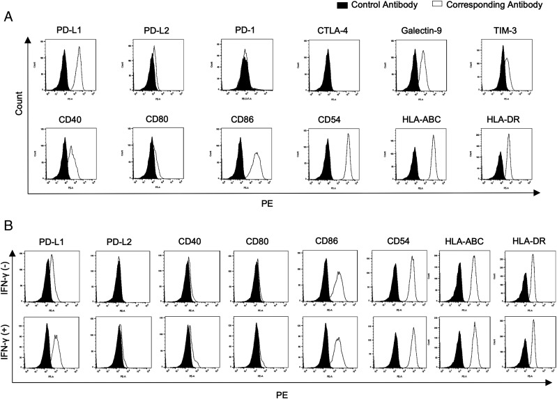
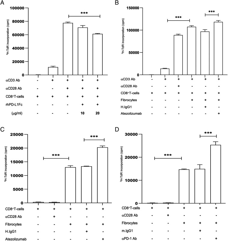
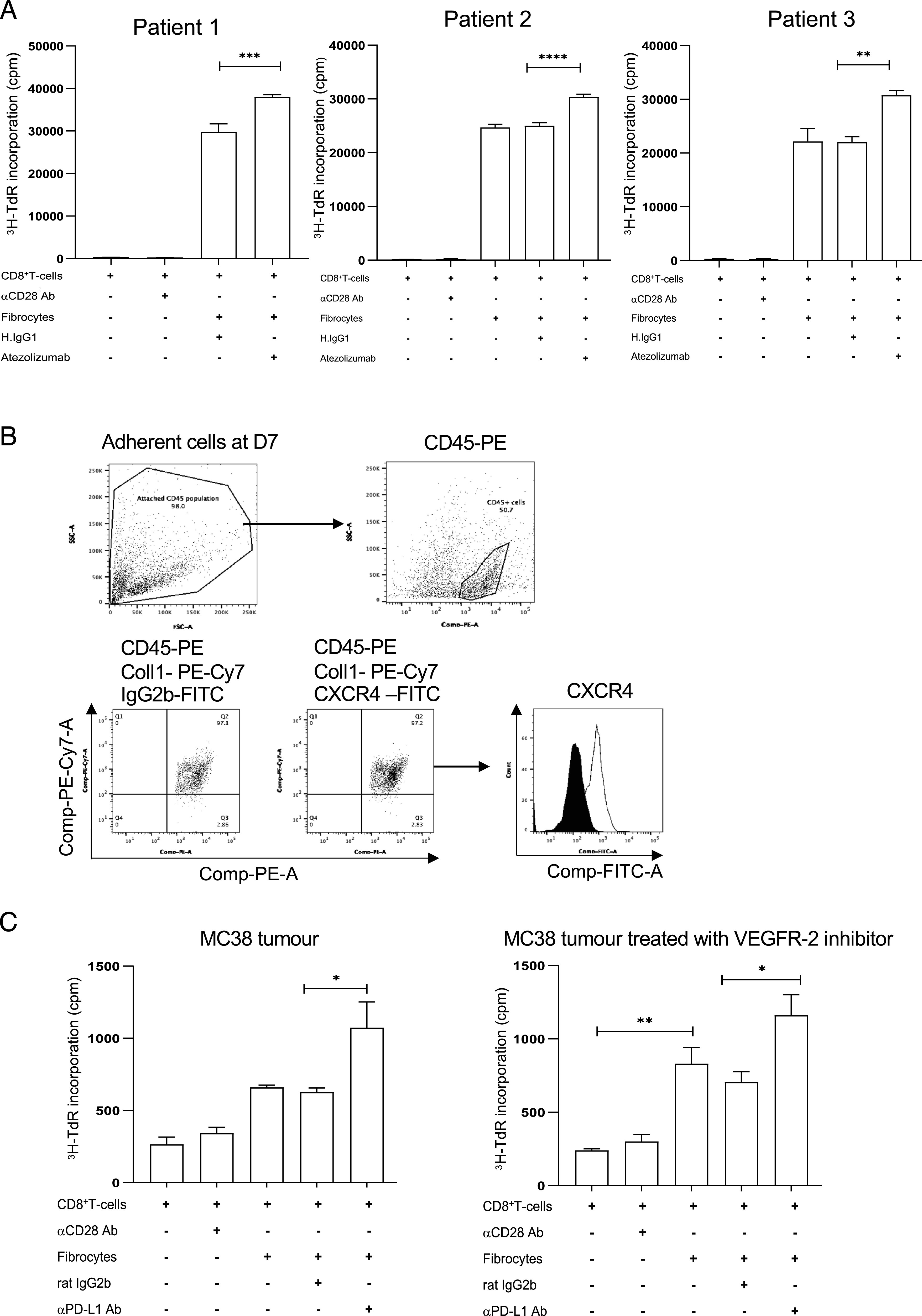
Fibrocytes, a distinct population of collagen-producing, monocyte-derived cells, are involved in wound healing as well as fibrotic diseases. Recently, fibrocytes have been revealed to play a role in the tumor microenvironment, particularly under antiangiogenic therapy. In addition, combination cancer immunotherapy with immune checkpoint inhibitor and antiangiogenic agents have been developed for various cancers in the clinical setting, although the immunological background is not clear. In the current study, we aimed to determine the function of fibrocytes in tumor immunity induced by immune checkpoint inhibitor therapy. Human and murine fibrocytes were generated from PBMCs and lungs, respectively. The expression of costimulatory and inhibitory molecules on fibrocytes was examined by flow cytometry. The stimulation of CD8+ T cells by fibrocytes was examined in MLRs with a 3H-thymidine incorporation assay. Fibrocytes expressed CD80low and CD86high as a costimulatory molecule, and expressed PD-L1high, but not PD-L2, as a coinhibitory molecule. Without any stimulation, fibrocytes strongly enhanced the proliferation of CD8+ T cells in mice and humans. Treatment with anti-CD86 and -CD54 Abs inhibited the growth of CD8+ T cells induced by fibrocytes. Anti-PD-L1 Ab further enhanced the proliferation of CD8+ T cells, even in the OVA-specific MLR with OT-1Rag-/- mice. Importantly, fibrocytes derived from PBMCs of patients with lung adenocarcinoma or murine MC38 tumors augmented the proliferation of CD8+ T cells with PD-L1 blockade. These results suggest that fibrocytes infiltrating tumor sites may play a role in the antitumor immunity mediated by CD8+ T cells when the activity is further enhanced by PD-L1/PD-1 blockade.


Conflict of interest statement

The authors have no financial conflicts of interest.

Figures

FIGURE 1.
FIGURE 1.
The cell surface expression of immune checkpoint molecules in fibrocytes and the effects of IFN-γ. A representative flow cytometric analysis of human PBMC-derived fibrocytes (A). The effects of IFN-γ on the expression of immune checkpoint molecules in human (B) fibrocytes. Data are representative of three independent experiments.
FIGURE 2.
FIGURE 2.
Fibrocytes stimulate the proliferation of CD8+ T cells through CD86 and CD54. (A) Human allogenic MLR with fibrocytes and DCs (n = 4). (B) The effects of blocking Abs against CD54, CD80, and CD86 in human allogeneic MLR stimulated with fibrocytes. (C) The effects of blocking Abs against CD54, CD80, and CD86 in human autologous MLR stimulated with fibrocytes and anti-CD3 Ab. ****p < 0.0001 by a one-way ANOVA, and data are shown as the mean ± SEM.
FIGURE 3.
FIGURE 3.
Blockade of the PD-1/PD-L1 pathway enhances the activation of CD8+ T cells by fibrocytes in human MLRs. (A) The effect of recombinant hPD-L1–Fc on the proliferation of CD8+ T cells activated by anti-CD3 and -CD28 Abs. (B) The effect of a humanized anti–hPD-L1 Ab, atezolizumab, on the proliferation of CD8+ T cells in autologous MLR stimulated with fibrocytes and anti-CD3 Ab. (C) The effect of a humanized anti–hPD-L1 Ab, atezolizumab, on the proliferation of CD8+ T cells in allogeneic MLR with fibrocytes. (D) The effect of anti–human PD-1 Ab on the proliferation of CD8+ T cells in allogeneic MLR with fibrocytes. Data are representative of three independent experiments. ***p < 0.001, by a one-way ANOVA, and data are shown as the mean ± SEM.
FIGURE 4.
FIGURE 4.
Comparison between fibrocytes and other APCs in allogeneic MLRs in human. Allogeneic MLRs with fibrocytes (A), DCs (B), and M1 (C) and M2 macrophages (D) from the same human donor. These data represent five or more repeated experiments and are shown as the mean ± SEM. **p < 0.01, by a one-way ANOVA.
FIGURE 5.
FIGURE 5.
The role of PD-L1 in Ag-specific MLR and cross-presentation by fibrocytes. (A) The effect of anti–murine PD-L1 Ab on the proliferation of CD8+ T cells from OT-1Rag−/− transgenic mice stimulated with fibrocytes pulsed with OVA peptide (SIINFEKL). (B) Murine MLR of CD8+ T cells from OT-1Rag−/− transgenic mice with fibrocytes or DCs pulsed with Ova protein. (C) Blocking of cross-presentation of fibrocytes with anti–MHC class I Ab. (D) The effect of anti–murine PD-L1 Ab on the proliferation of CD8+ T cells from OT-1Rag−/− transgenic mice with fibrocytes pulsed with OVA protein. Data are representative of three or more independent experiments. ****p < 0.0001, ***p < 0.001, by a one-way ANOVA, and data are shown as the mean ± SEM.
FIGURE 6.
FIGURE 6.
Blocking PD-L1 enhanced APC function of fibrocytes derived from patients with lung adenocarcinoma and murine tumors. (A) Allogeneic MLRs with CD8+ T cells derived from healthy donor and fibrocytes derived from patients with lung adenocarcinoma with atezolizumab (anti–PD-L1 Ab). (B) Flow-cytometric assessment of tumor-infiltrating fibrocytes derived from murine MC38 tumor. (C) Effect of anti–PD-L1 Ab on the proliferation of CD8+ T cells stimulated with fibrocytes in an allogeneic MLR. CD8+ T cells were harvested from spleen of BALB/c mice. Fibrocytes were harvested from MC38 tumors with or without treatment with VEGFR-2 inhibitor (semaxanib). ****p < 0.0001, ***p < 0.001, **p < 0.01, *p < 0.05, by a one-way ANOVA, and data are shown as the mean ± SEM.

Similar articles

Cited by

References

    1. Bucala, R., Spiegel L. A., Chesney J., Hogan M., Cerami A.. 1994. Circulating fibrocytes define a new leukocyte subpopulation that mediates tissue repair. Mol. Med. 1: 71–81. - PMC - PubMed
    1. Reilkoff, R. A., Bucala R., Herzog E. L.. 2011. Fibrocytes: emerging effector cells in chronic inflammation. Nat. Rev. Immunol. 11: 427–435. - PMC - PubMed
    1. Bellini, A., Mattoli S.. 2007. The role of the fibrocyte, a bone marrow-derived mesenchymal progenitor, in reactive and reparative fibroses. Lab. Invest. 87: 858–870. - PubMed
    1. Aono, Y., Kishi M., Yokota Y., Azuma M., Kinoshita K., Takezaki A., Sato S., Kawano H., Kishi J., Goto H., et al. . 2014. Role of platelet-derived growth factor/platelet-derived growth factor receptor axis in the trafficking of circulating fibrocytes in pulmonary fibrosis. Am. J. Respir. Cell Mol. Biol. 51: 793–801. - PubMed
    1. Gomperts, B. N., Strieter R. M.. 2007. Fibrocytes in lung disease. J. Leukoc. Biol. 82: 449–456. - PubMed

Publication types

MeSH terms