Case Reports
. 2021 Jan 27;8(2):e957.
doi: 10.1212/NXI.0000000000000957. Print 2021 Mar 4.

Presence of SARS-CoV-2 Transcripts in the Choroid Plexus of MS and Non-MS Patients With COVID-19

Affiliations
Case Reports

Presence of SARS-CoV-2 Transcripts in the Choroid Plexus of MS and Non-MS Patients With COVID-19

Vidmante Fuchs et al. Neurol Neuroimmunol Neuroinflamm. .
No abstract available


Figures

Figure 1
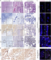
Figure 1. Histopathologic Assessment of MS COVID-19 Brain
(A–B) Macroscopically visible lesions were assessed by (A) H&E, LFB staining, and MBP, Iba1, CD45, and (B) dysferlin immunohistochemical evaluation. Given the lack of ongoing myelin phagocytosis presence of Iba1+ macrophages, all examined lesions were staged as chronic-inactive. Scale bar indicates 100 μm. (C) Assessing the presence and spatial distribution of severe acute respiratory syndrome coronavirus-2 (SARS-COV-2) transcripts by in situ hybridization in MS COVID-19 compared with non-COVID-19 tissue, we found presence of SARS-CoV-2 transcripts (pink arrows) in lung tissue, ependymal cells, and choroid plexus (CP) epithelial cells from MS-COVID-19 in comparable amount with non-MS COVID-19 CP epithelial cells with some coexpression of ACE2 (white arrows). Notably, SARS-CoV-2 transcripts were not present in MS lesions areas, suggesting CP epithelial cells as a key constraint of viral CNS entry. Arrows indicate positive cells. Left and right panel show different areas of the respective tissue section. Scale bar indicates 20 μm. COVID-19 = coronavirus disease 2019; H&E = hematoxylin and eosin; LFE = luxol fast blue; MBP = myelin basic protein; PV = periventricular; WM = white matter.

References

    1. Paterson RW, Brown RL, Benjamin L, et al. . The emerging spectrum of COVID-19 neurology: clinical, radiological and laboratory findings. Brain 2020;143:3104–3120. - PMC - PubMed
    1. Jaunmuktane Z, Mahadeva U, Green A, et al. . Microvascular injury and hypoxic damage: emerging neuropathological signatures in COVID-19. Acta Neuropathol 2020;140:397–400. - PMC - PubMed
    1. Deigendesch N, Sironi L, Kutza M, et al. . Correlates of critical illness-related encephalopathy predominate postmortem COVID-19 neuropathology. Acta Neuropathol 2020;140:583–586. - PMC - PubMed
    1. Sormani MP; Italian Study Group on C-iims. An Italian programme for COVID-19 infection in multiple sclerosis. Lancet Neurol 2020;19:481–482. - PMC - PubMed
    1. Haberman R, Axelrad J, Chen A, et al. . Covid-19 in immune-mediated inflammatory diseases—case series from New York. N Engl J Med 2020;383:85–88. - PMC - PubMed

Publication types