

Association of Dilated Perivascular Spaces With Cognitive Decline and Incident Dementia
- PMID: 33504642
- PMCID: PMC8032377
- DOI: 10.1212/WNL.0000000000011537
Association of Dilated Perivascular Spaces With Cognitive Decline and Incident Dementia
Abstract
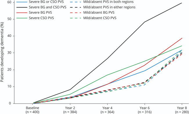
Objective: To determine whether severe perivascular space (PVS) dilation is associated with longitudinal cognitive decline and incident dementia over 4 and 8 years, respectively, we analyzed data from a prospective cohort study.
Methods: A total of 414 community-dwelling older adults aged 72-92 years were assessed at baseline and biennially for up to 8 years, with cognitive assessments, consensus dementia diagnoses, and 3T MRI. The numbers of PVS in 2 representative slices in the basal ganglia (BG) and centrum semiovale (CSO) were counted and severe PVS pathology defined as the top quartile. The effects of severe PVS pathology in either region or both regions and those with severe BG PVS and severe CSO PVS were examined. White matter hyperintensity volume, cerebral microbleed number, and lacune number were calculated.
Results: Participants with severe PVS pathology in both regions or in the CSO alone had greater decline in global cognition over 4 years, even after adjustment for the presence of other small vessel disease neuroimaging markers. The presence of severe PVS pathology in both regions was an independent predictor of dementia across 8 years (odds ratio 2.91, 95% confidence interval 1.43-5.95, p = 0.003). The presence of severe PVS pathology in all groups examined was associated with greater dementia risk at either year 4 or 6.
Conclusions: Severe PVS pathology is a marker for increased risk of cognitive decline and dementia, independent of other small vessel disease markers. The differential cognitive associations for BG and CSO PVS may represent differences in their underlying pathology.
© 2021 American Academy of Neurology.
Figures


References
-
- Doubal FN, MacLullich AM, Ferguson KJ, Dennis MS, Wardlaw JM. Enlarged perivascular spaces on MRI are a feature of cerebral small vessel disease. Stroke 2010;41:450–454. - PubMed
-
- Rouhl RP, van Oostenbrugge RJ, Knottnerus IL, Staals JE, Lodder J. Virchow-Robin spaces relate to cerebral small vessel disease severity. J Neurol 2008;255:692–696. - PubMed
-
- Ramirez J, Berezuk C, McNeely AA, Scott CJ, Gao F, Black SE. Visible Virchow-Robin spaces on magnetic resonance imaging of Alzheimer's disease patients and normal elderly from the Sunnybrook Dementia Study. J Alzheimers Dis 2015;43:415–424. - PubMed
-
- Zhu YC, Dufouil C, Soumare A, Mazoyer B, Chabriat H, Tzourio C. High degree of dilated Virchow-Robin spaces on MRI is associated with increased risk of dementia. J Alzheimers Dis 2010;22:663–672. - PubMed
Publication types
MeSH terms
LinkOut - more resources
Full Text Sources
Other Literature Sources
Medical
Research Materials