. 2021 Jan 27;6(1):e01136-20.
doi: 10.1128/mSphere.01136-20.

New Insights and Enhanced Human Norovirus Cultivation in Human Intestinal Enteroids

Affiliations

New Insights and Enhanced Human Norovirus Cultivation in Human Intestinal Enteroids

Khalil Ettayebi et al. mSphere. .

Abstract

Human noroviruses (HuNoVs) are the leading cause of epidemic and sporadic acute gastroenteritis worldwide. We previously demonstrated human intestinal stem cell-derived enteroids (HIEs) support cultivation of several HuNoV strains. However, HIEs did not support virus replication from every HuNoV-positive stool sample, which led us to test and optimize new medium conditions, identify characteristics of stool samples that allow replication, and evaluate consistency of replication over time. Optimization of our HIE-HuNoV culture system has shown the following: (i) a new HIE culture medium made with conditioned medium from a single cell line and commercial media promotes robust replication of HuNoV strains that replicated poorly in HIEs grown in our original culture medium made with conditioned media from 3 separate cell lines; (ii) GI.1, 11 GII genotypes (GII.1, GII.2, GII.3, GII.4, GII.6, GII.7, GII.8, GII.12, GII.13, GII.14, and GII.17), and six GII.4 variants can be cultivated in HIEs; (iii) successful replication is more likely with virus in stools with higher virus titers; (iv) GII.4_Sydney_2012 virus replication was reproducible over 3 years; and (v) HuNoV infection is restricted to the small intestine, based on replication of two viral strains in duodenal and ileal HIEs, but not colonoids, from two susceptible donors. These results improve the HIE culture system for HuNoV replication. Use of HIEs by several laboratories worldwide to study the molecular mechanisms that regulate HuNoV replication confirms the usefulness of this culture system, and our optimized methods for virus replication will advance the development of effective therapies and methods for virus control.IMPORTANCE Human noroviruses (HuNoVs) are highly contagious and cause acute and sporadic diarrheal illness in all age groups. In addition, chronic infections occur in immunocompromised cancer and transplant patients. These viruses are antigenically and genetically diverse, and there are strain-specific differences in binding to cellular attachment factors. In addition, new discoveries are being made on strain-specific differences in virus entry and replication and the epithelial cell response to infection in human intestinal enteroids. Human intestinal enteroids are a biologically relevant model to study HuNoVs; however, not all strains can be cultivated at this time. A complete understanding of HuNoV biology thus requires cultivation conditions that will allow the replication of multiple strains. We report optimization of HuNoV cultivation in human intestinal enteroid cultures to increase the numbers of cultivatable strains and the magnitude of replication, which is critical for testing antivirals, neutralizing antibodies, and methods of virus inactivation.

Keywords: human intestinal enteroids; human norovirus; virus replication.


Figures

FIG 1
FIG 1
Successful replication is more likely with virus from stools with higher virus titers (A) but not affected by patient age (B). (A) Replication of GII.4 strains plotted by virus titer. Dashed vertical line indicates the GII.4_Sydney_2012 ID50 determined previously (12). Data points indicate stools from patients under (green) and over (blue) 2 years of age. (B) Replication of the same set of GII.4 strains plotted by patient age. Purple, stool titer > GII.4_Sydney_2012 ID50. Orange, stool titer < GII.4_Sydney_2012 ID50. Replication less than 0.5 log10 was assigned a value of 0. Dashed lines show the detection 0.5-log10 threshold.
FIG 2
FIG 2
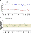
Replication of GII.4_2012_Sydney in HIEs plated in BCM medium is reproducible over time. (A) Virus replication of GII.4_Sydney_2012 HuNoV, included as a positive control in different experiments throughout 3+ years to assess the reproducibility of viral infection in J2 HIE monolayers infected with 9 × 105 GEs/well in BCM medium, was determined at 1 hpi and 24 hpi. (B) Fold change at 24 hpi compared to 1 hpi. The mean log10 increase at 24 hpi versus 1 hpi was 2.25 (n = 80). Error bars denote standard deviation from 6 wells in each experiment.
FIG 3
FIG 3
Improved HuNoV replication in different jejunal HIE cultures plated as monolayers in INT medium. (A) Schematic design of HIE culture maintenance and monolayer seeding prior to infection (“p” and “d” notations refer to proliferation and differentiation, respectively). (B and C) GII.4_Sydney_2012 (9 × 105 GEs/well) (B) and GII.3 (4.3 × 105 GEs/well) (C) virus replication was evaluated in four jejunal (J2, J3, J6, and J11) HIE lines plated in either BCM or INT medium. Each experiment was performed twice, and compiled data are presented. Error bars denote standard deviation (n = 12). Values above the bars indicate log10 (fold change) replication difference in INT versus BCM medium at 24 hpi. Significance was determined using Student’s t test (***, P value < 0.001).
FIG 4
FIG 4
HuNoV replication in HIE cultures from different intestinal segments (duodenum, ileum, and colon) from two independent donors (104 and 109). HIEs were plated in BCMp or INTp medium (see schematic design in Fig. 3A). After differentiation, monolayers were infected with GII.4_Sydney_2012 (9 × 105 GEs/well) (A and C) or GII.3 (4.3 × 105 GEs/well) (B and D). Compiled data from two experiments are presented. Error bars denote standard deviation (n = 12), and each data bar represents the mean for six wells of inoculated HIE monolayers. Values on the bars indicate log10 (fold change) replication difference in INT versus BCM medium at 24 hpi. Significance was determined using Student’s t test (***, P value < 0.001; n.s., not significant).
FIG 5
FIG 5
Replication of GII.4_2012_Sydney in HIEs plated in Intesticult medium is reproducible and less variable over 1 year. (A) Virus replication of GII.4_2012_Sydney HuNoV, included as a positive control in different experiments throughout 1 year (2019) to assess the reproducibility of viral infection in J2 HIE monolayers inoculated with 9 × 105 GEs/well in INT medium, was determined at 1 hpi and 24 hpi. (B) Fold change at 24 hpi compared to 1 hpi. The mean log10 increase at 24 hpi versus 1 hpi was 2.66 (n = 23). Error bars denote standard deviation (n = 6).
FIG 6
FIG 6
Replication of different GII HuNoV genotypes is improved when HIEs are plated in INT medium. J2 HIEs were initially maintained as 3D-HIEs in BCMp or L-WRN proliferation medium and then plated and differentiated for 5 days in the indicated medium (A, experimental design). Monolayers were inoculated with GII.4_Sydney_2012 (9 × 105 GEs/well) (B), GII.3 (4.3 × 105 GEs/well) (C), GII.17_1295-44 (4.6 × 105 GEs/well) (D), or GII.6_TCH13-106 (3.3 × 105 GEs/well) (E) diluted in CMGF[−] with 500 μM GCDCA. After 1 hpi, monolayers were washed and cultured in the indicated BCM or INT differentiation medium (+ 500 μM GCDCA). Values represent log10 difference in viral growth between conditions [(Δ24hpi-1hpi in INT) – (Δ24hpi-1hpi in BCM)]. Error bars denote standard deviation (n = 6). Asterisks indicate significant difference between conditions. ***, P value < 0.001; **, P < 0.01; *, P < 0.05; n.s., not significant.
FIG 7
FIG 7
Medium composition can improve HuNoV replication. J2 HIEs were propagated in INT or L-WRN medium prior to seeding them into monolayers. Monolayers were prepared from INT-3D- or L-WRN-3D-HIEs, proliferated for 1 day, and then differentiated for 5 days in the indicated medium (A, experimental design). They were inoculated with GII.4_Sydney_2012 (9 × 105 GEs/well) (B), GII.3 (4.3 × 105 GEs/well) (C), GII.17_1295-44 (4.6 × 105 GEs/well) (D), or GII.6_TCH13-106 (3.3 × 105 GEs/well) (E) diluted in CMGF[−] with 500 μM GCDCA. After 1 hpi, monolayers were washed twice and cultured in the indicated differentiation medium (+ 500 μM GCDCA). Values above bars represent log10 difference in viral growth between conditions. Error bars denote standard deviation (n = 6). Asterisks indicate significant difference between conditions.

References

    1. Chhabra P, de Graaf M, Parra GI, Chan MC, Green K, Martella V, Wang Q, White PA, Katayama K, Vennema H, Koopmans MPG, Vinjé J. 2019. Updated classification of norovirus genogroups and genotypes. J Gen Virol 100:1393–1406. doi:10.1099/jgv.0.001318. - DOI - PMC - PubMed
    1. Ahmed SM, Hall AJ, Robinson AE, Verhoef L, Premkumar P, Parashar UD, Koopmans M, Lopman BA. 2014. Global prevalence of norovirus in cases of gastroenteritis: a systematic review and meta-analysis. Lancet Infect Dis 14:725–730. doi:10.1016/S1473-3099(14)70767-4. - DOI - PMC - PubMed
    1. Hall AJ, Lopman BA, Payne DC, Patel MM, Gastañaduy PA, Vinjé J, Parashar UD. 2013. Norovirus disease in the United States. Emerg Infect Dis 19:1198–1205. doi:10.3201/eid1908.130465. - DOI - PMC - PubMed
    1. Atmar RL, Ramani S, Estes MK. 2018. Human noroviruses: recent advances in a 50-year history. Curr Opin Infect Dis 31:422–432. doi:10.1097/QCO.0000000000000476. - DOI - PubMed
    1. Bartsch SM, Lopman BA, Ozawa S, Hall AJ, Lee BY. 2016. Global economic burden of norovirus gastroenteritis. PLoS One 11:e0151219. doi:10.1371/journal.pone.0151219. - DOI - PMC - PubMed

Publication types