. 2021 Jan 27;12(1):610.
doi: 10.1038/s41467-020-20586-2.

Endophilin A2 deficiency protects rodents from autoimmune arthritis by modulating T cell activation

Affiliations

Endophilin A2 deficiency protects rodents from autoimmune arthritis by modulating T cell activation

Ulrika Norin et al. Nat Commun. .

Abstract

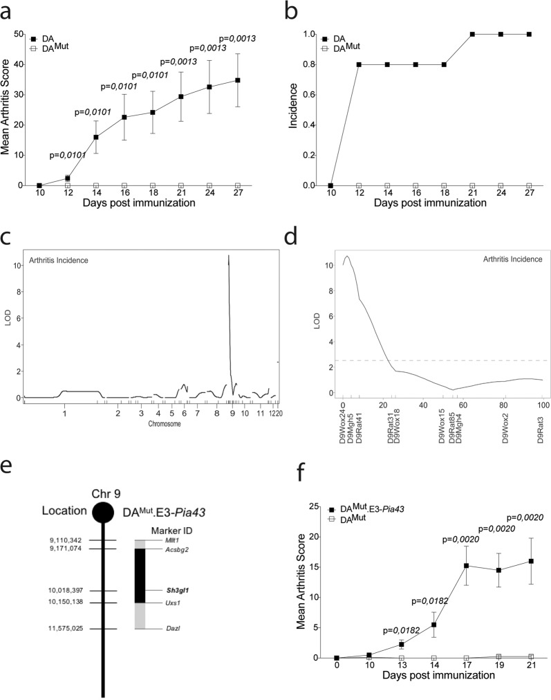
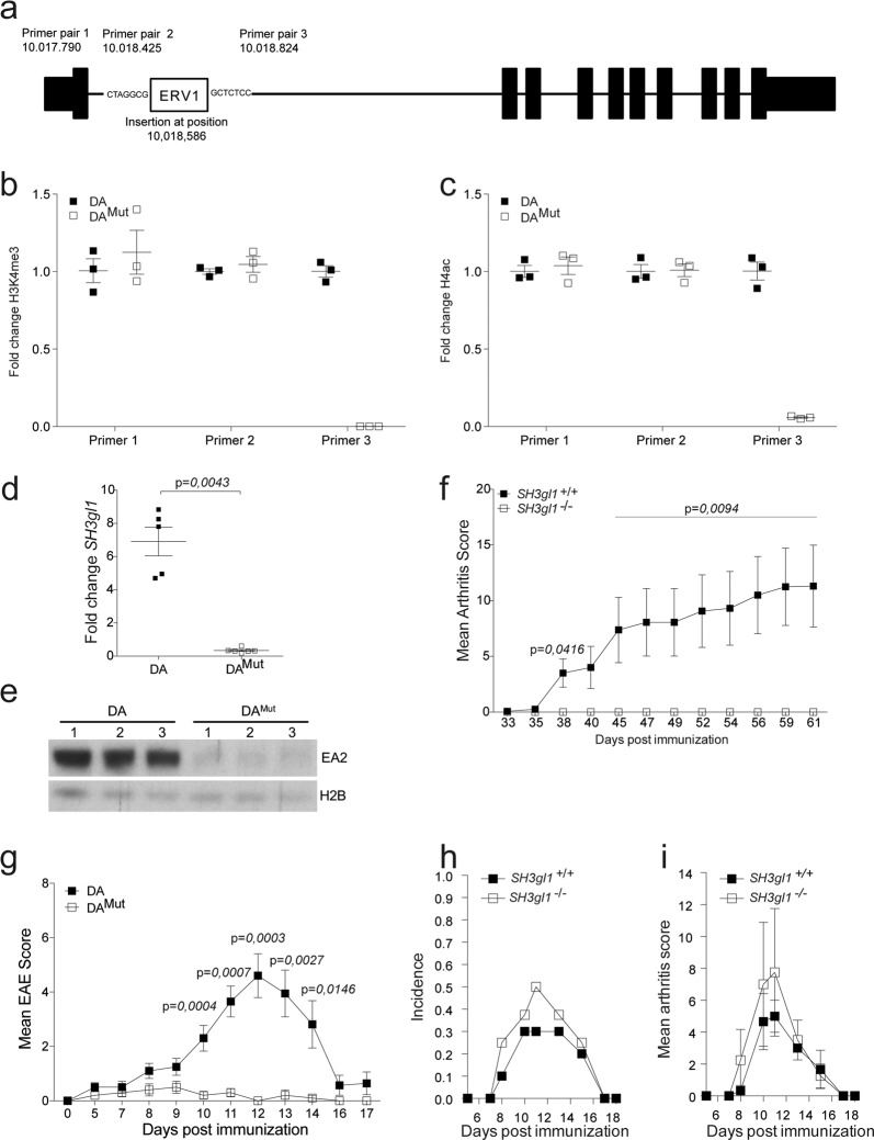
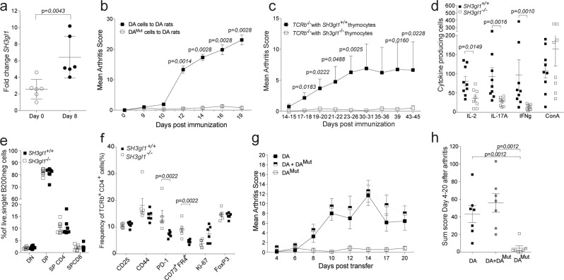
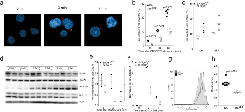
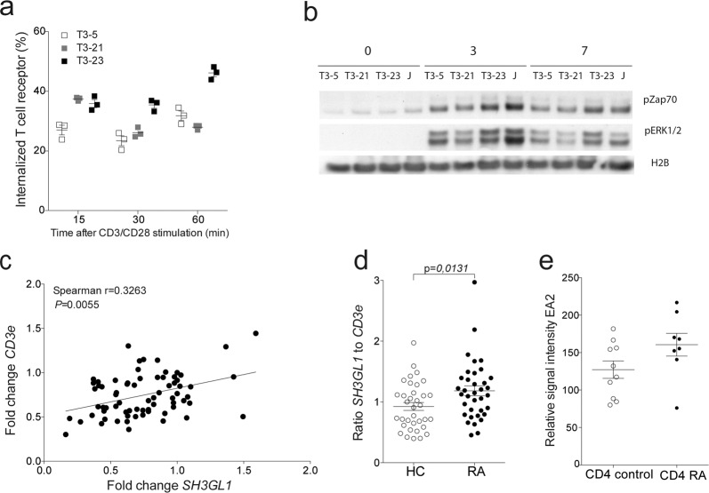
The introduction of the CTLA-4 recombinant fusion protein has demonstrated therapeutic effects by selectively modulating T-cell activation in rheumatoid arthritis. Here we show, using a forward genetic approach, that a mutation in the SH3gl1 gene encoding the endocytic protein Endophilin A2 is associated with the development of arthritis in rodents. Defective expression of SH3gl1 affects T cell effector functions and alters the activation threshold of autoreactive T cells, thereby leading to complete protection from chronic autoimmune inflammatory disease in both mice and rats. We further show that SH3GL1 regulates human T cell signaling and T cell receptor internalization, and its expression is upregulated in rheumatoid arthritis patients. Collectively our data identify SH3GL1 as a key regulator of T cell activation, and as a potential target for treatment of autoimmune diseases.


Conflict of interest statement

The authors declare no competing interests.

Figures

Fig. 1
Fig. 1. Arthritis regulating loci in the DAMut rat is restricted to a 2-Mb region on chromosome 9.
a Mean arthritis score after pristane-induced arthritis in 5 DA and 7 DAMut rats. The arthritis data has been reproduced three times with the same results. b Incidence of arthritis after pristane-induced arthritis in DA and DAMut rats. c LOD score plot for incidence of PIA in 51 (E3.DA-Pia457x DAMut) x DAMut rats of all genotyped chromosomes. d LOD score plot for incidence of PIA in (E3.DA-Pia457 × DAMut) DAMut rats on chromosome 9, broken line at 2.63 indicate significant linkage e Schematic figure of the DAMut.E3-Pia43 congenic fragment on chromosome 9 with location of the SH3gl1 gene. Microsatellite markers in the Acsbg2 and Uxs1 genes indicate inner border of congenic fragment and markers Mllt1 and Dazl indicate outer borders. f Mean arthritis score after pristane-induced arthritis in 8 DAMut and 4 DAMut E3-Pia43 heterozygote littermates. Arthritis has been reproduced in several different congenic fragments with the same results. Non-parametrical Mann–Whitney U test was used for statistical evaluation of data. Data are presented as mean with error bars indicating ± SEM with each dot representing an individual value.
Fig. 2
Fig. 2. Endophilin A2 deficiency protects against autoimmunity in rodents.
a Schematic picture of the SH3gl1 gene with the location of the inserted ERVI long-term repeat element and location of the primer pairs used for quantification of the H3K4me3 and H4Ac levels. b Fold change of H3K4me3 levels in 3 DA and 3 DAMut naïve aged matched rats before (Primer pair 1 and 2) and after (Primer pair 3) the insertion in the SH3gl1 gene. c Fold change of H4Ac levels in 3 DA and 3 DAMut naïve aged matched rats before (Primer pair 1 and 2) and after (Primer pair 3) the insertion in the SH3gl1 gene. d Fold change of the SH3gl1 gene expression in PBMCs from 5 DA and 6 DAMut naïve aged matched rats. Relative fold change calculated to one DAMut reference sample. e Western blot analysis of the expression of the Endophilin A2 protein in brain samples of three DA and DAMut rats. Histone 2B was used as loading control. f Mean arthritis score after collagen-induced arthritis in 9 SH3gl1 knockouts and 16 wild-type littermates. Arthritis data has been reproduced twice with the same results. g Mean EAE score after spinal cord homogenate induced EAE in 10 DA and 10 DAMut rats. h Incidence of collagen-antibody induced arthritis in 10 SH3gl1−/− and 10 SH3gl1+/+ wild-type littermates. i Mean arthritis score of sick mice from above experiment after collagen-antibody induced arthritis in 4 SH3gl1 knockouts and three wild-type littermates. Non-parametrical Mann–Whitney U test was used for statistical evaluation of data. Data are presented as mean with error bars indicating ±SEM with each dot representing an individual value.
Fig. 3
Fig. 3. T cells regulate Endophilin A2 mediated protection against arthritis.
a Expression of SH3gl1 in sorted T cells from lymph nodes from DA rats at Day 0 and Day 8 after pristane immunization. b Mean arthritis score after adoptive transfer of pristane-primed DA and DAMut cells to 9 respectively 4 naïve DA recipients. The adoptive transfer experiment has been reproduced a total of three times with the same results. c Mean arthritis score after GPI-induced arthritis in 17 and 19 TCRβ knockout recipients that had received either SH3gl1−/− or SH3gl1+/+ thymocytes respectively, 7 days prior to disease induction. Data is pooled from three individual experiments. d Number of antigen specific IL-2, IL-17A and IFNg producing T cells after recall stimulation with GPI peptide and IL-2 producing cells after ConA stimulation day 44 after GPI-induced arthritis. Data from 9 TCRβ knockout mice reconstituted with wild-type T cells and 8 TCRβ knockout mice reconstituted SH3gl1 T cells. Recall experiment has been repeated twice in SH3gl1−/− mice. e Frequency of double negative (DN), double positive (DP), single positive CD4 (SP CD4), and single positive CD8 (SP CD8) thymocytes in 8 SH3gl1−/− and 8 SH3gl1+/+ mice. Data has been reproduced twice. f Frequency of CD25, CD44, PD-1, CD73+FR4+, Ki-67, and FoxP3 positive cells in CD4+TCRb+ cells from inguinal lymph nodes ten days after GPI-peptide immunization from 6 SH3gl1−/− and 6 SH3gl1+/+ mice. g Mean arthritis score in naïve DA recipients after adoptive transfer of pristane-primed DAMut cells only, DA cells only or DA cells in addition with DAMut cells, 7 rats per group. h Arthritis sum score of the DA recipients above after adoptive transfer of pristane-primed DA cells only or addition with DAMut cells, 7 rats per group. Non-parametrical Mann–Whitney U test was used for statistical evaluation of data. Data are presented as mean with error bars indicating ±SEM with each dot representing an individual value.
Fig. 4
Fig. 4. Endophilin A2 co-localizes with the TCR upon activation and regulates TCR internalization and signaling.
a EA2 co-localization with the TCR in Jurkat cells after anti-CD3/CD28 stimulation determined by proximity-ligase assay and visualized as TexasRed+ spots using confocal imaging. b Percentage of internalized TCR in T cells from 5 DA and 5 DAMut rats after anti-CD3/CD28 stimulation. Data has been reproduced two times. c Percentage of internalized TCR in CD4+CD3+ T cells from three SH3gl1−/− and SH3gl1+/+ mice stimulated with anti-CD3/CD28 for 30 min in the presence or absence of Brefeldin A at 1.25 μg/ml. Experiment has been repeated twice with the same results. d Representative Western blot analysis from two experiments of phosphorylated and unphosphorylated ZAP70 and ERK1/2 in T cells from two SH3gl1−/− and SH3gl1+/+ mice stimulated with anti-CD3/CD28 for 0, 3, and 7 min. Histone 2B was used as loading control. e Normalized OD values of phosphorylated Zap70 compared to unphosphorylated Zap70 from two SH3gl1−/− and SH3gl1+/+ mice. f Normalized OD values of phosphorylated ERK1/2 compared to unphosphorylated ERK1/2 from two SH3gl1−/− and SH3gl1+/+ mice. g Flow cytometry blot showing in vitro cell proliferation of DAMut and DA T cells 72 h after anti-CD3/CD28 stimulation. h Division index of sorted T cells from 7 DA and 5 DAMut rats after 72 h of anti-CD3/CD28 stimulation. Non-parametrical Mann–Whitney U test was used for statistical evaluation of data. Data are presented as mean with error bars indicating ± SEM with each dot representing an individual value.
Fig. 5
Fig. 5. EA2 has a conserved function in human T cells and is upregulated in RA patients.
a TCR internalization in SH3GL1 CRISPR knockout Jurkat cells (T3-5 and T3-21) and wild-type control (T3-23). b Representative western blot analysis from two experiments of phosphorylated ZAP70 and ERK1/2 in T cells from in SH3GL1 CRISPR knockout (T3-5 and T3-21), wild-type control (T3-23), and Jurkat (J) cells with anti-CD3/CD28 for 0, 3, and 7 min. Histone 2B was used as loading control. c Correlation of CD3e and SH3GL1 gene expression in whole blood of healthy controls and RA patients. Each circle represents one individual. d Normalized SH3GL1 gene expression to CD3e gene expression in whole blood of 35 healthy controls and 36 RA patients, randomly selected from the cohort used in c. Each circle represents one individual. e SH3GL1 gene expression in sorted CD4+ T cells from 10 healthy controls and 8 RA patients (data accessible at NCBI GEO database, accession GSE4588). Non-parametrical Mann–Whitney U test was used for statistical evaluation of data. Data are presented as mean with error bars indicating ±SEM with each dot representing an individual value.

References

    1. Bos WH, van de Stadt LA, Sohrabian A, Rönnelid J, van Schaardenburg D. Development of anti-citrullinated protein antibody and rheumatoid factor isotypes prior to the onset of rheumatoid arthritis. Arthritis Res. Ther. 2014;16:405. doi: 10.1186/ar4511. - DOI - PMC - PubMed
    1. Siebert S, Tsoukas A, Robertson J, McInnes I. Cytokines as therapeutic targets in rheumatoid arthritis and other inflammatory diseases. Pharmacol. Rev. 2015;67:280–309. doi: 10.1124/pr.114.009639. - DOI - PubMed
    1. Okada Y, et al. Genetics of rheumatoid arthritis contributes to biology and drug discovery. Nature. 2014;506:376–381. doi: 10.1038/nature12873. - DOI - PMC - PubMed
    1. Van Boxel JA, Paget SA. Predominantly T-cell infiltrate in rheumatoid synovial membranes. N. Engl. J. Med. 1975;293:517–520. doi: 10.1056/NEJM197509112931101. - DOI - PubMed
    1. Ranges GE, Sriram S, Cooper SM. Prevention of type II collagen-induced arthritis by in vivo treatment with anti-L3T4. J. Exp. Med. 1985;162:1105–1110. doi: 10.1084/jem.162.3.1105. - DOI - PMC - PubMed

Publication types

MeSH terms

Substances