. 2021 Jan 27;7(1):9.
doi: 10.1038/s41522-020-00177-1.

Reduction of alternative electron acceptors drives biofilm formation in Shewanella algae

Affiliations

Reduction of alternative electron acceptors drives biofilm formation in Shewanella algae

Alberto J Martín-Rodríguez et al. NPJ Biofilms Microbiomes. .

Abstract

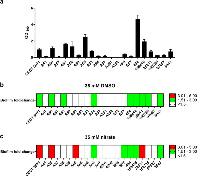
Shewanella spp. possess a broad respiratory versatility, which contributes to the occupation of hypoxic and anoxic environmental or host-associated niches. Here, we observe a strain-specific induction of biofilm formation in response to supplementation with the anaerobic electron acceptors dimethyl sulfoxide (DMSO) and nitrate in a panel of Shewanella algae isolates. The respiration-driven biofilm response is not observed in DMSO and nitrate reductase deletion mutants of the type strain S. algae CECT 5071, and can be restored upon complementation with the corresponding reductase operon(s) but not by an operon containing a catalytically inactive nitrate reductase. The distinct transcriptional changes, proportional to the effect of these compounds on biofilm formation, include cyclic di-GMP (c-di-GMP) turnover genes. In support, ectopic expression of the c-di-GMP phosphodiesterase YhjH of Salmonella Typhimurium but not its catalytically inactive variant decreased biofilm formation. The respiration-dependent biofilm response of S. algae may permit differential colonization of environmental or host niches.


Conflict of interest statement

The authors declare no competing interests.

Figures

Fig. 1
Fig. 1. Biofilm formation response of S. algae strains in the absence or presence of supplemented alternative electron acceptors.
a Biofilm formation for 21 S. algae strains in MB medium in the absence of added electron acceptors. b Heatmap representing the fold-change in biofilm formation with respect to the corresponding non-supplemented control upon addition of 35 mM DMSO. c Heatmap representing the fold-change in biofilm formation with respect to the corresponding non-supplemented control upon addition of 35 mM sodium nitrate. Data represent the average of two biological replicates with six technical replicates each.
Fig. 2
Fig. 2. Nitrate and DMSO reduction capacity of S. algae strains.
a Biofilm induction is not related to the ability of the strains to utilize the supplemented compound, as indicated by a qualitative nitrate reduction test performed for all 21 strains, showing nitrite production as indicated by the formation of a pink complex. Uninoculated MB medium supplemented with 35 mM nitrate was used as blank. b Evolutionary history of DmsA orthologs inferred by using the Maximum Likelihood method and Whelan and Goldman model. Bootstrap support values are indicated in the nodes of the phylogenetic reconstruction.
Fig. 3
Fig. 3. Electron acceptors promote strain-specific biofilm formation of S. algae.
a Experimental set-up to assess biofilm formation by S. algae. Alternative electron acceptors (AEAs) were incorporated into ultra-pure agarose, from which they are progressively released. b Visualization of AEA release from agarose. Shown is a recipient with agarose containing cobalt (II) nitrate that is overlaid with water. c Surface colonization patterns of S. algae CECT 5071 and S. algae A291 on agarose plugs containing either no AEAs or 35 mM of the selected AEAs DMSO or sodium nitrate visualized by Confocal Laser Scanning Microscopy after staining with the BacLight viability kit. Representative images are shown.
Fig. 4
Fig. 4. Disruption of terminal reductase activity abrogates the biofilm response.
a Schematic of the DMSO reductase encoding dmsEFABGH operon of Shewanella algae. b DMSO reduction of the WT strain and dmsB in-frame deletion mutant (****P < 0.0001, two-tailed unpaired t test) as evidenced by measuring DMS production of three independent cultures supplemented with 35 mM DMSO. c Biofilm formation of S. algae CECT 5071 WT and ∆dmsB in the absence or presence of DMSO (****P < 0.0001, two-tailed unpaired t test). Data represent the average and SD of three biological replicates with seven technical replicates each. d Schematic of the periplasmic nitrate reductase NAP-α encoding operon napEDABC and NAP-β encoding operon napDAGHB of S. algae. e Nitrate reduction by the single mutants ∆napABC) and ∆napA), and the double mutant ∆napABCnapA (αβ) as determined by nitrite accumulation with respect to the WT strain upon supplementation with 35 mM nitrate (****P < 0.0001; one-way ANOVA followed by Dunnett’s post-hoc test; ND no nitrite production detected). Data represent the average and SD of three biological replicates with four technical replicates per sample. f Biofilm formation of S. algae CECT 5071 WT and nitrate reduction mutants in the absence or presence of sodium nitrate (****P < 0.0001, one-way ANOVA followed by Dunnett’s post-hoc test).
Fig. 5
Fig. 5. Ectopic expression of terminal reductases restores catalytic activity and biofilm formation.
a Restoration of DMSO reductase activity as determined by DMS production in the ∆dmsB mutant upon overexpression of the dmsEFAGHB operon (average ± SD, n = 3) in comparison to the WT and ∆dmsB strains harboring the empty expression plasmid pSRK-Km (****P < 0.0001; one-way ANOVA followed by Tukey’s post-hoc test). b Biofilm formation of the complemented ∆dmsB mutant and the corresponding WT and ∆dmsB empty pSRK-Km vector controls. Data represent the average and SD of three biological replicates with seven technical replicates per sample. Statistical significance of the differences was determined by one-way ANOVA followed by Tukey’s post-hoc test (***P < 0.001, ns not significant). c Biofilm formation patterns on the walls and bottom of the wells of S. algae WT pSRK-Km, S. algaedmsB pSRK-Km, and complemented S. algaedmsB pSRK-Km-dmsEFABGH as visually assessed by CV staining. Note that biofilm formation differences on the bottom of the wells by the WT harboring the empty plasmid pSRK-Km and complemented mutant with respect to the ∆dmsB mutant harboring the empty expression vector are not apparent from the quantitative determinations shown on b because of the vector effect. d Nitrate reductase activity in complemented α, β, and αβ mutants as determined by nitrite production, as well as in a catalytic napEDABC mutant in which three of the four cysteines of the 4Fe-4S cluster of NapA had been replaced by serine. Data represent the average and SD of three biological replicates with four technical replicates per sample. Statistical significance of the differences was determined by one-way ANOVA followed by Tukey’s post-hoc test (*P < 0.05, **P < 0.01, ***P < 0.001, ****P < 0.0001, ND no nitrite production detected). e Biofilm formation of complemented nitrate reduction mutants and the corresponding WT and nitrate reduction null strains harboring the empty pSRK-Km plasmid, and the catalytic mutant. Data represent the average and SD of three biological replicates with seven technical replicates each. Statistical significance of the differences was determined by one-way ANOVA followed by Tukey’s post-hoc test.
Fig. 6
Fig. 6. Transcriptomic profiles of S. algae CECT 5071 static cultures supplemented with DMSO or nitrate.
Venn diagrams of (a) differentially expressed genes (P-adj < 0.05) in both treatments, (b) genes with upregulated transcript levels and (c) genes with downregulated transcript levels. Volcano plots showing alterations in gene transcript levels in static cultures supplemented with 35 mM DMSO (d) and nitrate (e). In red are the genes that are significantly differentially regulated (P-adj < 0.05) with a fold-change value below 2 or above 0.5. In orange are the rest of differentially expressed genes. Black indicates genes not significantly differentially regulated. COG assignation of differentially expressed genes in S. algae CECT 5071 upon addition of 35 mM DMSO (f) or sodium nitrate (g) versus control cultures without electron acceptor addition. COG categories: [A] RNA processing and modification; [B] Chromatin structure and dynamics; [C] Energy production and conversion; [D] Cell cycle control, cell division, chromosome partitioning; [E] Amino acid transport and metabolism; [F] Nucleotide transport and metabolism; [G] Carbohydrate transport and metabolism; [H] Coenzyme transport and metabolism; [I] Lipid transport and metabolism; [J] Translation, ribosomal structure and biogenesis; [K] Transcription; [L] Replication, recombination and repair; [M] Cell wall/membrane/envelope biogenesis; [N] Cell motility; [O] Post-translational modification, protein turnover, and chaperones; [P] Inorganic ion transport and metabolism; [Q] Secondary metabolites biosynthesis, transport, and catabolism; [R] General function prediction only; [S] Function unknown; [T] Signal transduction mechanisms; [U] Intracellular trafficking, secretion, and vesicular transport; [V] Defense mechanisms; [W] Extracellular structures; [Y] Nuclear structure; and [Z] Cytoskeleton.
Fig. 7
Fig. 7. Putative role of c-di-GMP signaling on AEA-induced biofilm formation.
a Heatmap showing transcriptional changes (log2 fold-change) of genes encoding putative c-di-GMP turnover proteins upon DMSO or nitrate supplementation. b Biofilm formation of S. algae CECT 5071 WT harboring the empty plasmid pBBR1MCS-2 (denoted as VC for “vector control”) or expressing the PDE from S. Typhimurium YhjH or the catalytically inactive derivative YhjH E136A cloned in this plasmid in the absence or in the presence of 35 mM DMSO or nitrate. Data represent the average and SD of three biological replicates with seven technical replicates per sample. Statistical significance of the differences was determined by one-way ANOVA followed by Tukey’s post-hoc test (****P < 0.0001, ns not significant).

Similar articles

Cited by

References

    1. Nealson, K. H. & Scott, J. Ecophysiology of the genus Shewanella. In The Prokaryotes. 1133–1151 (Springer New York, 2006).
    1. Hau HH, Gralnick JA. Ecology and biotechnology of the genus Shewanella. Annu. Rev. Microbiol. 2007;61:237–258. doi: 10.1146/annurev.micro.61.080706.093257. - DOI - PubMed
    1. Kotloski NJ, Gralnick JA. Flavin electron shuttles dominate extracellular electron transfer by Shewanella oneidensis. MBio. 2013;4:e00553–12. doi: 10.1128/mBio.00553-12. - DOI - PMC - PubMed
    1. Martín-Rodríguez AJ, Martín-Pujol O, Artiles-Campelo F, Bolaños-Rivero M, Römling U. Shewanella spp. infections in Gran Canaria, Spain:retrospective analysis of 31 cases and a literature review. JMM Case Rep. 2017;4:e005131. doi: 10.1099/jmmcr.0.005131. - DOI - PMC - PubMed
    1. Martín-Rodríguez AJ, Suárez-Mesa A, Artiles-Campelo F, Römling U, Hernández M. Multilocus sequence typing of Shewanella algae isolates identifies disease-causing Shewanella chilikensis strain 6I4. FEMS Microbiol. Ecol. 2019;95:fiy210. - PubMed

Publication types

MeSH terms

Supplementary concepts