. 2021 Jan 27;11(1):2291.
doi: 10.1038/s41598-021-81638-1.

Altered high-density lipoprotein composition and functions during severe COVID-19

Affiliations

Altered high-density lipoprotein composition and functions during severe COVID-19

Floran Begue et al. Sci Rep. .

Abstract

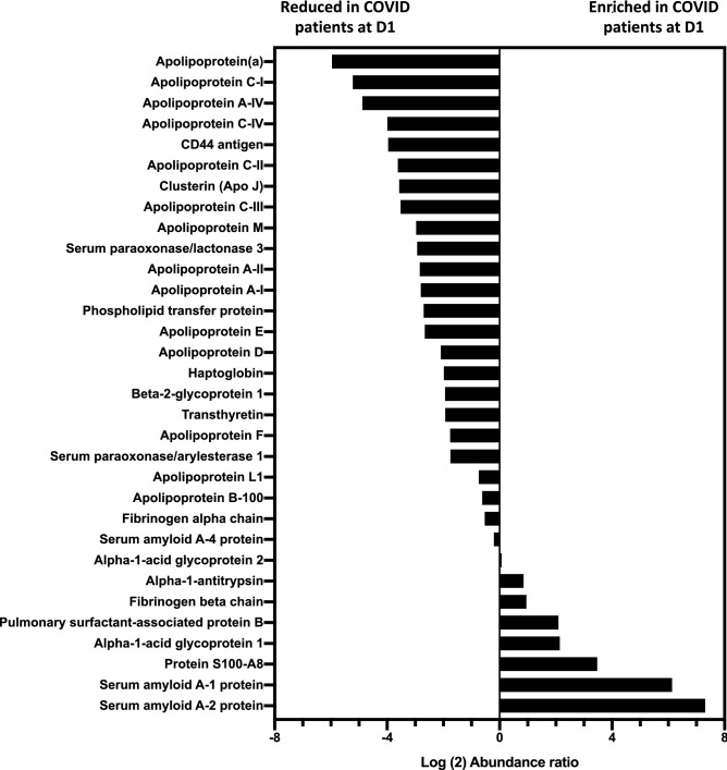
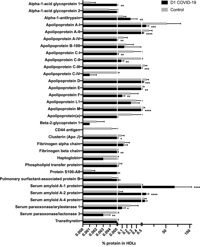
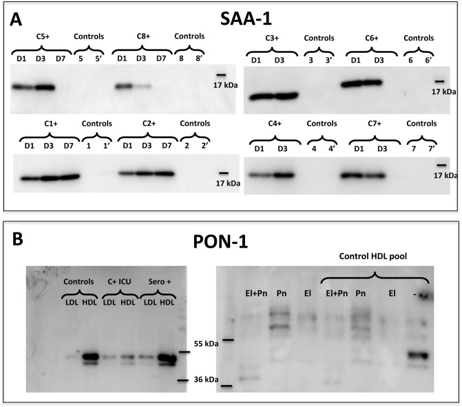
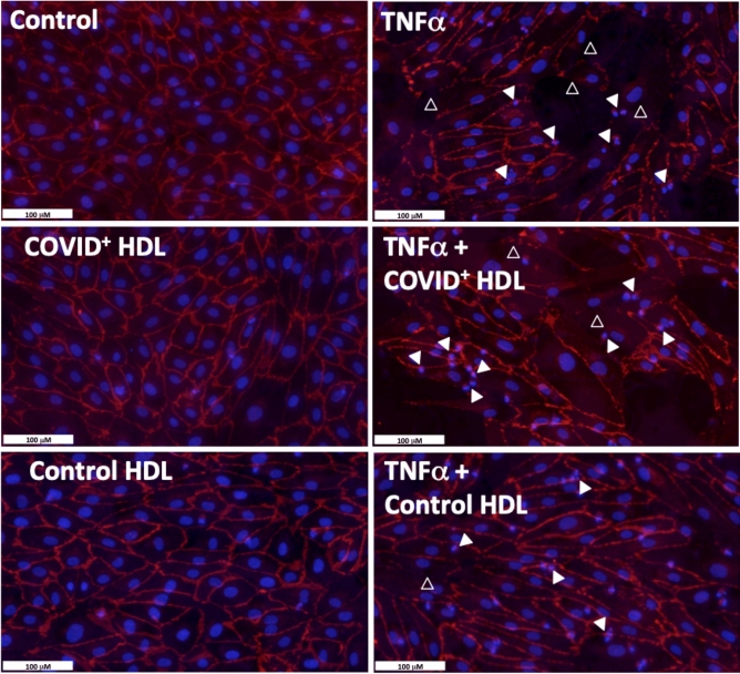
Coronavirus disease 2019 (COVID-19) pandemic is affecting millions of patients worldwide. The consequences of initial exposure to SARS-CoV-2 go beyond pulmonary damage, with a particular impact on lipid metabolism. Decreased levels in HDL-C were reported in COVID-19 patients. Since HDL particles display antioxidant, anti-inflammatory and potential anti-infectious properties, we aimed at characterizing HDL proteome and functionality during COVID-19 relative to healthy subjects. HDLs were isolated from plasma of 8 severe COVID-19 patients sampled at admission to intensive care unit (Day 1, D1) at D3 and D7, and from 16 sex- and age-matched healthy subjects. Proteomic analysis was performed by LC-MS/MS. The relative amounts of proteins identified in HDLs were compared between COVID-19 and controls. apolipoprotein A-I and paraoxonase 1 were confirmed by Western-blot analysis to be less abundant in COVID-19 versus controls, whereas serum amyloid A and alpha-1 antitrypsin were higher. HDLs from patients were less protective in endothelial cells stiumalted by TNFα (permeability, VE-cadherin disorganization and apoptosis). In these conditions, HDL inhibition of apoptosis was blunted in COVID-19 relative to controls. In conclusion, we show major changes in HDL proteome and decreased functionality in severe COVID-19 patients.


Conflict of interest statement

The authors declare no competing interests.

Figures

Figure 1
Figure 1
Relative abundance of proteins identified in HDLs isolated from of the plasma of control subjects (n = 15) and COVID-19 patients (n = 8) at admission in ICU (D1). The relative abundance of proteins in HDLs from controls and D1 COVID-19 patients was expressed as the Log2 Abundance ratio (D1 COVID/Control), as described in the “Methods” section.
Figure 2
Figure 2
Percentage of protein abundance in HDLs from controls and COVID-19 patients. HDLs were isolated from the plasma of 15 controls and 8 patients with COVID-19 at admission (D1). Bar graphs represent the percentage of intensity ± SD of the two groups. Unpaired t-test was used. *p < 0.0332, **p < 0.0021, ***p < 0.0002 and ****p < 0.0001 as compared to the control group.
Figure 3
Figure 3
Western blot analysis for detection of SAA and PON-1 in HDL isolated from COVID-19 patients and controls. HDLs (3 μg protein /lane) were immunoblotted for SAA (serum amyloid A) (A) and PON-1 (paraoxonase 1) (B). (A) HDLs isolated from patients and controls are presented individually (C1+ = ICU-COVID-19 positive patient #1, C2+ for patient #2, etc.) at day 1, 3 and 7 after admission (D1, 3 and 7). Controls 1 and 1′ correspond to HDLs isolated from healthy subjects sex- and age-matched to patient #1. (B) Pools of LDLs and HDLs were blotted for PON-1 (left panel) after isolation from non-infected healthy subjects ("Controls"), COVID-19 positive patients in intensive care unit ("C+ICU") (n = 8) and SARS-CoV-2 seropositive cured caregivers ("sero+", sampled > 15 days after symptoms, n = 7). A pool of control HDLs containing PON-1 was incubated with elastase (El, 20 μg/mL), plasmin (Pn, 500 μg/mL) or both proteases (El + Pn) for 2 h at 37 °C. Enzymes alone (without HDLs) treated in the same experimental conditions were loaded as control. Uncropped gels are presented in Supplementary information file.
Figure 4
Figure 4
Real-time monitoring of HUVEC barrier dysfunction in response to TNFα+/−HDLs. Human umbilical vein endothelial cells (25,000 cells/well) were seeded on gelatin-coated xCELLigence 16 well E-plates. HDL were isolated from plasma of 16 controls and 8 D1 COVID ICU patients by ultracentrifugation and pooled into two groups: “Control HDL" and “COVID+ HDL”. The cell index was recorded continuously until confluence, when a plateau was reached [indicated cell growth in (A)]. Cells were then serum-deprived (- Fetal Calf Serum, FCS) for 6 h and then stimulated with 30 ng/mL TNFα+/−HDLs from COVID-19 patients or controls at 0.2 mg/mL. Cell indexes were normalized after stimulation [as indicated by the vertical line, in (A), 30 min. after the stimulation]. A magnification showing TNFα, TNFα + COVID + HDL and TNFα + control HDL is presented in (B). Representative results from 2 independent experiments. HDLs isolated from each patient and matched controls (2 controls/patients) were analyzed separately (Supplementary Fig. 4A).
Figure 5
Figure 5
Effect of HDLs from controls and COVID-19 (C+) subjects on HUVEC apoptosis cells after stimulation with 30 ng/mL TNFα for 24 h. HDL were isolated from plasma of 16 controls and 8 D1 COVID-19 subjects by ultracentrifugation and pooled into two groups respectively “Control HDL" and “COVID+ HDL” Upper panel: immunostaining for VE-Cadherin (red) and nuclear staining with DAPI (blue). Empty white arrowheads show VE-Cadherin re-organization. Plain white arrowheads show examples of apoptotic nuclei (condensed and/or fragmented).
Figure 6
Figure 6
TUNEL and DAPI staining- Quantification of apoptotic nuclei. In TNFα-treated HUVECs (30 ng/mL 24 h), dual staining shows a good correlation between morphological apoptotic features determined by nuclear condensation and fragmentation (DAPI in blue, A, C) and DNA fragmentation (green, B, C). Bar graph (D) represents the means ± SD of apoptotic nuclei count in 5 fields (between 700 and 900 nuclei counted) of a representative experiment. *p = 0.0332 (Control vs TNFα). **p = 0.002 (Control vs TNFα + COVID+ HDL). #p = 0.03 (TNFα + control HDL vs TNFα + COVID+ HDL).

Similar articles

  • Dysfunctional HDL in acute stroke.
    Ortiz-Munoz G, Couret D, Lapergue B, Bruckert E, Meseguer E, Amarenco P, Meilhac O. Ortiz-Munoz G, et al. Atherosclerosis. 2016 Oct;253:75-80. doi: 10.1016/j.atherosclerosis.2016.08.035. Epub 2016 Aug 26. Atherosclerosis. 2016. PMID: 27591364
  • HDL proteome remodeling associates with COVID-19 severity.
    Souza Junior DR, Silva ARM, Rosa-Fernandes L, Reis LR, Alexandria G, Bhosale SD, Ghilardi FR, Dalçóquio TF, Bertolin AJ, Nicolau JC, Marinho CRF, Wrenger C, Larsen MR, Siciliano RF, Di Mascio P, Palmisano G, Ronsein GE. Souza Junior DR, et al. J Clin Lipidol. 2021 Nov-Dec;15(6):796-804. doi: 10.1016/j.jacl.2021.10.005. Epub 2021 Oct 31. J Clin Lipidol. 2021. PMID: 34802985 Free PMC article.
  • HDL proteome and apolipoproteins concentrations in severe ICU COVID-19 patients.
    Begue F, Veeren B, Rondeau P, Florence AM, Jamard S, Montravers P, Tanaka S, Meilhac O; French COVID Study Group. Begue F, et al. Lipids Health Dis. 2025 Jan 31;24(1):32. doi: 10.1186/s12944-024-02381-w. Lipids Health Dis. 2025. PMID: 39891286 Free PMC article.
  • High-density lipoproteins and COVID-19: preparing the next pandemic.
    Apalama ML, Begue F, Tanaka S, Cournot M, Couret D, Meilhac O, Pokeerbux MR. Apalama ML, et al. J Lipid Res. 2025 May;66(5):100779. doi: 10.1016/j.jlr.2025.100779. Epub 2025 Mar 14. J Lipid Res. 2025. PMID: 40090619 Free PMC article. Review.
  • COVIDomics: The Proteomic and Metabolomic Signatures of COVID-19.
    Costanzo M, Caterino M, Fedele R, Cevenini A, Pontillo M, Barra L, Ruoppolo M. Costanzo M, et al. Int J Mol Sci. 2022 Feb 22;23(5):2414. doi: 10.3390/ijms23052414. Int J Mol Sci. 2022. PMID: 35269564 Free PMC article. Review.

Cited by

References

    1. Hirano T, Murakami M. COVID-19: A new virus, but a familiar receptor and cytokine release syndrome. Immunity. 2020;52:731–733. doi: 10.1016/j.immuni.2020.04.003. - DOI - PMC - PubMed
    1. Fan J, et al. Letter to the Editor: Low-density lipoprotein is a potential predictor of poor prognosis in patients with coronavirus disease 2019. Metabolism. 2020;107:154243. doi: 10.1016/j.metabol.2020.154243. - DOI - PMC - PubMed
    1. Wang D, et al. Correlation analysis between disease severity and clinical and biochemical characteristics of 143 cases of COVID-19 in Wuhan, China: A descriptive study. BMC Infect. Dis. 2020;20:519. doi: 10.1186/s12879-020-05242-w. - DOI - PMC - PubMed
    1. Hu X, Chen D, Wu L, He G, Ye W. Declined serum high density lipoprotein cholesterol is associated with the severity of COVID-19 infection. Clin. Chim. Acta. 2020;510:105–110. doi: 10.1016/j.cca.2020.07.015. - DOI - PMC - PubMed
    1. Tanaka S, et al. Lipoprotein concentrations over time in the intensive care unit COVID-19 patients: Results from the ApoCOVID study. PLoS ONE. 2020;15:e0239573. doi: 10.1371/journal.pone.0239573. - DOI - PMC - PubMed

Publication types

MeSH terms