. 2021 Jan 27;11(1):2363.
doi: 10.1038/s41598-021-81900-6.

Detection of colorectal cancer in urine using DNA methylation analysis

Affiliations

Detection of colorectal cancer in urine using DNA methylation analysis

S Bach et al. Sci Rep. .

Abstract

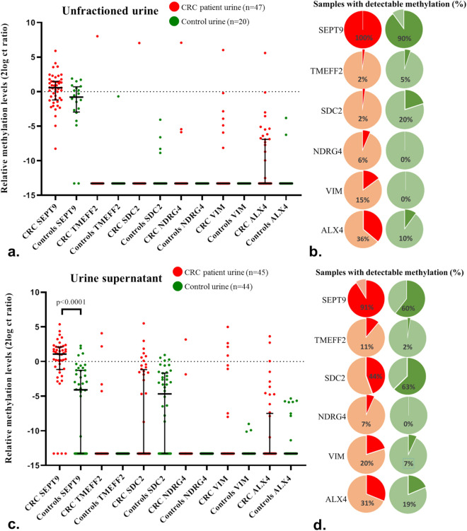
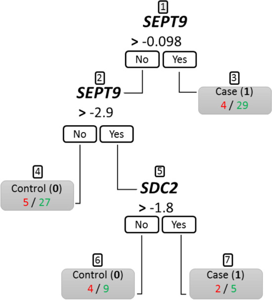
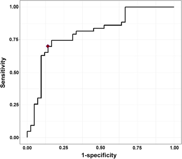
Colorectal cancer (CRC) is the second leading cause for cancer-related death globally. Clinically, there is an urgent need for non-invasive CRC detection. This study assessed the feasibility of CRC detection by analysis of tumor-derived methylated DNA fragments in urine. Urine samples, including both unfractioned and supernatant urine fractions, of 92 CRC patients and 63 healthy volunteers were analyzed for DNA methylation levels of 6 CRC-associated markers (SEPT9, TMEFF2, SDC2, NDRG4, VIM and ALX4). Optimal marker panels were determined by two statistical approaches. Methylation levels of SEPT9 were significantly increased in urine supernatant of CRC patients compared to controls (p < 0.0001). Methylation analysis in unfractioned urine appeared inaccurate. Following multivariate logistic regression and classification and regression tree analysis, a marker panel consisting of SEPT9 and SDC2 was able to detect up to 70% of CRC cases in urine supernatant at 86% specificity. First evidence is provided for CRC detection in urine by SEPT9 methylation analysis, which combined with SDC2 allows for an optimal differentiation between CRC patients and controls. Urine therefore provides a promising liquid biopsy for non-invasive CRC detection.


Conflict of interest statement

R.D.M.S has a minority stake in Self-screen B.V., a spin-off company of VU University Medical Center Amsterdam. R.D.M.S., S.B., I.B. and G.K. are named inventors on patent application(s) related to the detection of cancer DNA in urine. All other authors have no conflict of interest to declare.

Figures

Figure 1
Figure 1
Methylation levels (a + c) and detection rates (b + d) in both unfractioned urine and urine supernatant samples. (a, c) Shows methylation detection of SEPT9, TMEFF2, SDC2, NDRG4, VIM and ALX4 in unfractioned and supernatant urine samples of CRC patients and controls. Data are shown as the median with interquartile range of 2log converted Ct ratios. In  b, c, detection rates of markers in unfractioned urines and supernatant samples of CRC patients and controls are depicted, meaning the percentage of samples scoring a CtMARKER value below 45.
Figure 2
Figure 2
SEPT9 methylation levels in urine supernatant per CRC stage. Samples of patient with stage IV CRC have been further stratified in patients with and without the primary tumor still being present. Furthermore, methylation levels in patients with solely intraperitoneal metastases (n = 15) and patients diagnosed with stage IV due to liver metastases (n = 5) are shown. Data are shown as the median with interquartile range of 2log converted Ct ratios.
Figure 3
Figure 3
The CART decision tree. The boxes depict the decision nodes. Based on SEPT9 or SDC2 methylation values, samples are classified as a case (1) or control (0). The numbers below the node represent urine samples that are classified incorrectly (red) or correctly (green) according to the classification of 0 or 1. Node numbers are indicated above by 1 to 7.
Figure 4
Figure 4
The ROC curve for classification of supernatant urine samples using multivariate logistics regression (MLR) model with marker SEPT9 and interaction of markers SEPT9 and SDC2. The red diamond indicates the maximal Youden’s index.
Figure 5
Figure 5
The performance of both generated models and their accuracy in detection of CRC in supernatant urine. The figure shows the classification of CRC samples and controls for both MLR and CART models. At the bottom, performance specifics for both models are depicted.

References

    1. Bray F, et al. Global cancer statistics 2018: GLOBOCAN estimates of incidence and mortality worldwide for 36 cancers in 185 countries. CA Cancer J. Clin. 2018;68:394–424. doi: 10.3322/caac.21492. - DOI - PubMed
    1. Siegel RL, et al. Colorectal cancer statistics, 2017. CA Cancer J. Clin. 2017;67:177–193. doi: 10.3322/caac.21395. - DOI - PubMed
    1. Bach S, et al. Circulating tumor DNA analysis: clinical implications for colorectal cancer patients. A Systematic Review. JNCI Cancer Spect. 2019 doi: 10.1093/jncics/pkz042. - DOI - PMC - PubMed
    1. Issa IA, Noureddine M. Colorectal cancer screening: an updated review of the available options. World J. Gastroenterol. 2017;23:5086–5096. doi: 10.3748/wjg.v23.i28.5086. - DOI - PMC - PubMed
    1. Rex DK, et al. Colorectal cancer screening: recommendations for physicians and patients from the U.S. Multi-Society Task Force on Colorectal Cancer. Am. J. Gastroenterol. 2017;112:1016–1030. doi: 10.1038/ajg.2017.174. - DOI - PubMed

Publication types