. 2021 Jan 27;4(1):123.
doi: 10.1038/s42003-020-01633-6.

Elucidating the tunability of binding behavior for the MERS-CoV macro domain with NAD metabolites

Affiliations

Elucidating the tunability of binding behavior for the MERS-CoV macro domain with NAD metabolites

Meng-Hsuan Lin et al. Commun Biol. .

Abstract

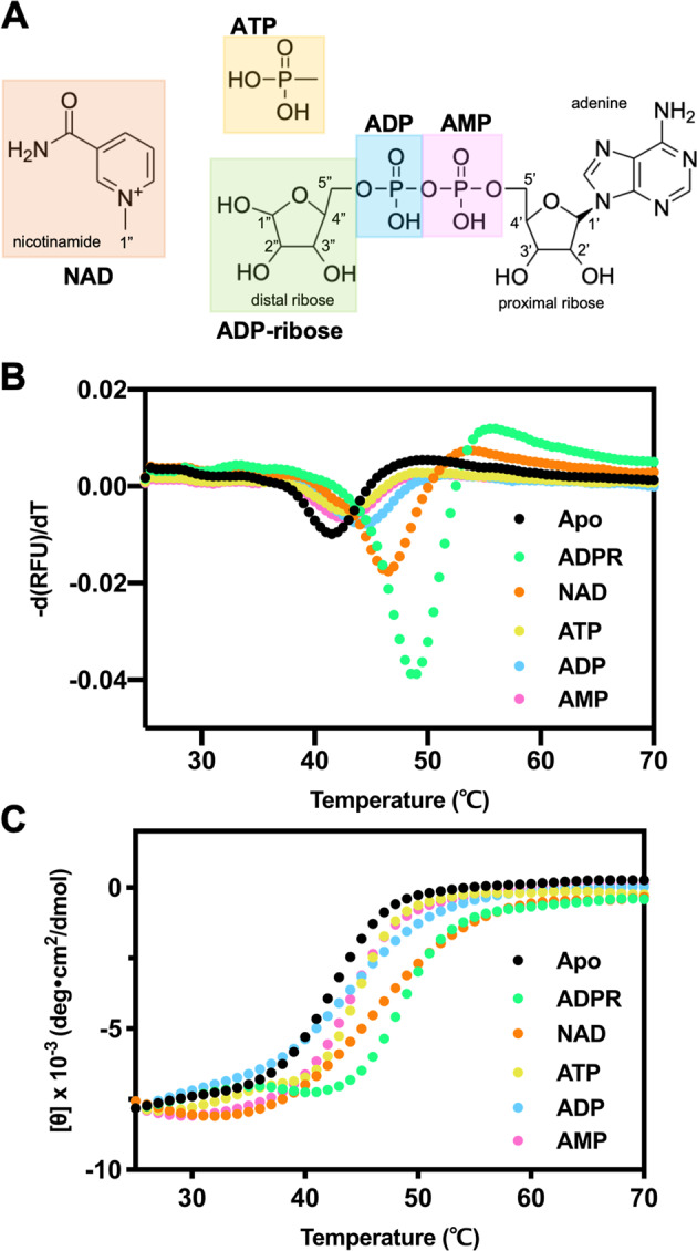
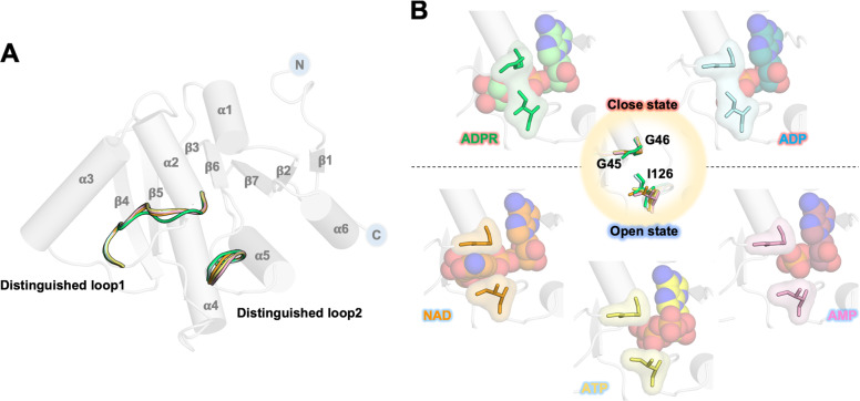
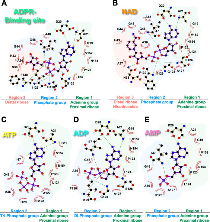
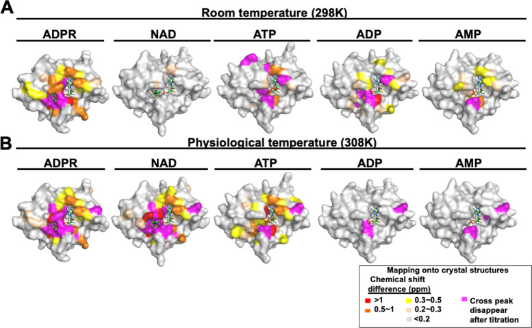
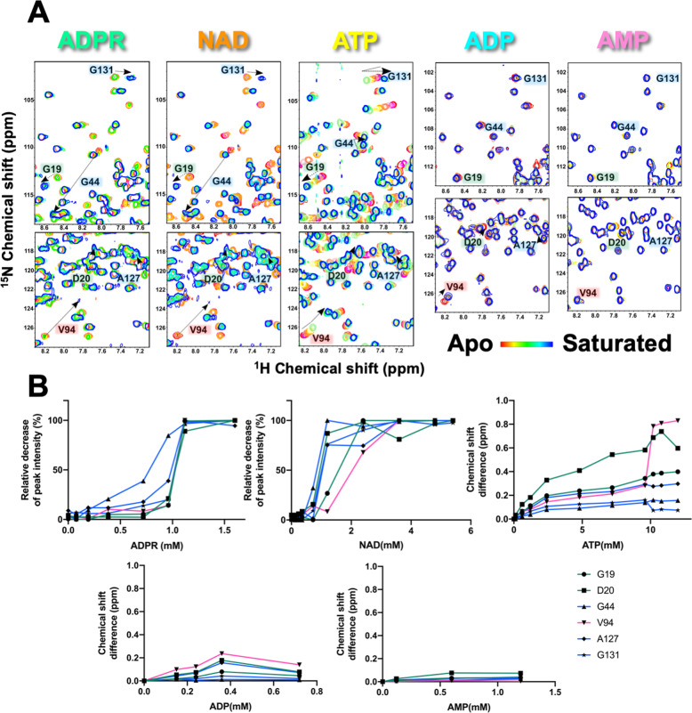
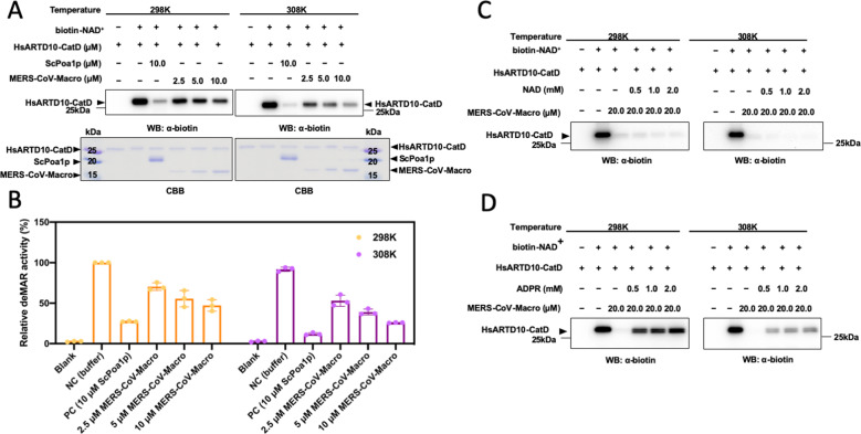
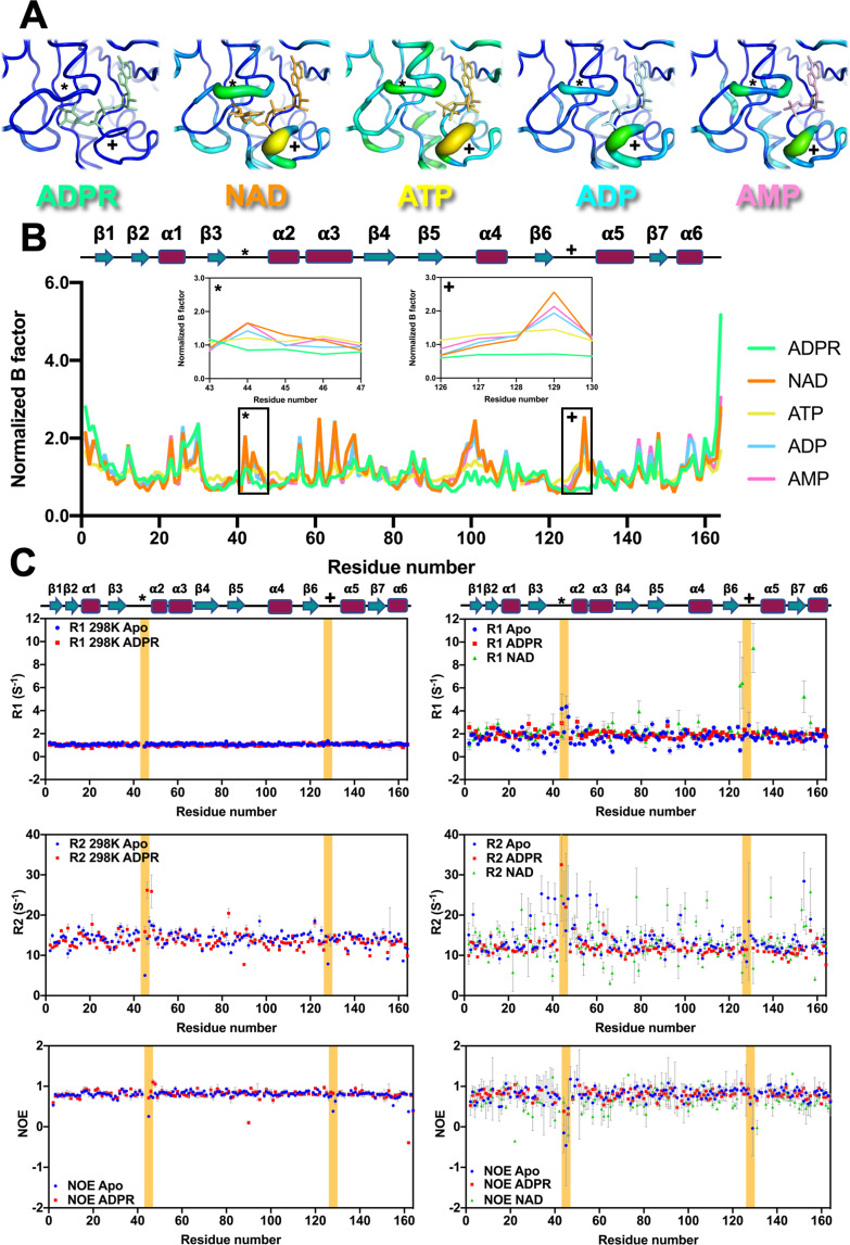
The macro domain is an ADP-ribose (ADPR) binding module, which is considered to act as a sensor to recognize nicotinamide adenine dinucleotide (NAD) metabolites, including poly ADPR (PAR) and other small molecules. The recognition of macro domains with various ligands is important for a variety of biological functions involved in NAD metabolism, including DNA repair, chromatin remodeling, maintenance of genomic stability, and response to viral infection. Nevertheless, how the macro domain binds to moieties with such structural obstacles using a simple cleft remains a puzzle. We systematically investigated the Middle East respiratory syndrome-coronavirus (MERS-CoV) macro domain for its ligand selectivity and binding properties by structural and biophysical approaches. Of interest, NAD, which is considered not to interact with macro domains, was co-crystallized with the MERS-CoV macro domain. Further studies at physiological temperature revealed that NAD has similar binding ability with ADPR because of the accommodation of the thermal-tunable binding pocket. This study provides the biochemical and structural bases of the detailed ligand-binding mode of the MERS-CoV macro domain. In addition, our observation of enhanced binding affinity of the MERS-CoV macro domain to NAD at physiological temperature highlights the need for further study to reveal the biological functions.


Conflict of interest statement

The authors declare no competing interests.

Figures

Fig. 1
Fig. 1. Evaluation of MERS-CoV macro domain binding with NAD metabolites by Tm values.
A Chemical structures of NAD metabolites tested in this study. Melting temperatures of 10 μM MERS-CoV macro domain in complex with 1 mM NAD metabolites were determined by B thermal shift assays and C circular dichroism. MERS-CoV macro domain in the apo form, ADPR-bound, NAD-bound, ATP-bound, ADP-bound, and AMP-bound form are labeled in black circles, green squares, orange triangles, yellow triangles, blue diamonds, and pink circles, respectively.
Fig. 2
Fig. 2. Two divergences between the five structures of the MERS-CoV macro domain in complex with different NAD metabolites.
A Two distinguished loops in protein structures complexed with ADPR, NAD, ATP, ADP, and AMP are represented by ribbon models colored in light green, orange, yellow, cyan, and light pink, respectively. B The closed/open states of the ligand-binding pocket result from the orientation of residues, G45, G46, and I126, located on the two distinguished loops.
Fig. 3
Fig. 3. Ligplot diagrams for NAD metabolites coordinated with the MERS-CoV macro domain.
Components of A ADPR-binding, B NAD-binding, C ATP-binding, D ADP-binding, and E AMP-binding sites are shown. According to the chemical structure of each ligand, the binding sites are classed in several regions: region 1 in green, region 2 in marine, and region3 in salmon. Covalent bonds of ligands are in purple, covalent bonds of amino acids are in brown, and hydrogen bonds are in green dashed lines. Protein residues in hydrophobic contacts with NAD metabolites are represented by eyelash symbols.
Fig. 4
Fig. 4. Surface mapping of the MERS-CoV macro domain with significant chemical shift changes observed in NMR perturbation at 298 K (A) and 308 K (B).
Surface colors of the MERS-CoV macro domain in complex with various NAD metabolites according to chemical shift difference upon ligand titration.
Fig. 5
Fig. 5. Chemical shift perturbations of residues in three regions of ligand binding site at 308 K.
A Overlays of the 1H-15N HSQC spectra of residues in three regions of the ligand-binding site of the MERS-CoV macro domain alone and a series of NAD metabolites titrations. B The decrease in intensity of cross-peaks and the chemical shift difference of residues selected are plotted against the concentration of titrated NAD metabolites.
Fig. 6
Fig. 6. De-MARylation of the MERS-CoV macro domain at different temperatures.
A The deMAR enzyme activity of the MERS-CoV macro domain. Auto-mono-ADP-ribosylated human ARTD 10 catalytic domain (HsARTD10-CatD) was arranged using biotin-NAD+ as a substrate. Budding yeast macro domain Poa1p (ScPoa1p) is a positive control. Varying amounts of the MERS-CoV macro domain (from 2.5 to 10.0 μM) mixing with biotin labeled MAR-HsARTD10-CatD at 298 and 308 K were resolved by 15% acrylamide gel. The level of mono-ADP-ribosylation was detected by western blot assay using antibody anti-biotin. B The relative intensity of the bands corresponding to MAR-remained after the reactions of the MERS-CoV macro domain at different temperatures. Independent experiments n = 3. The deMAR enzyme activity of the MERS-CoV macro domain with the presence of C NAD and D ADPR, respectively. Auto-mono-ADP-ribosylated human ARTD 10 catalytic domain (HsARTD10-CatD) was arranged using biotin-NAD+ as a substrate. Varying amounts of C NAD or D ADPR mixing with 20.0 μM MERS-CoV macro domain and biotin-labeled MAR-HsARTD10-CatD at 298 K and 308 K were resolved by 15% acrylamide gel. The level of mono-ADP-ribosylation was detected by western blot assay using antibody antibiotin.
Fig. 7
Fig. 7. A tunable ligand-binding site of the MERS-CoV macro domain.
A Ligand binding site of MERS-CoV macro domain complexed with different NAD metabolites colored according to the B-factor putty script distributed with PyMOL. Distinguished loops are labeled by symbols, stars (*), and crosses (+). B Comparison of normalized B factor among various NAD metabolite-bound forms of the MERS-CoV macro domain. The flexibility difference at the distinguished loops of the ligand-binding site (*, +) is shown by insert plots of normalized B factor vs. residue number. C NMR relaxation parameters, R1, R2, NOE, of the MERS-CoV macro domain apo form, ADPR-complex, and NAD-complex form labeled by blue circles, red squares, and green triangles, respectively. The left and right panel show relaxation parameters determined at 298 K and 308 K, respectively. Parts of distinguished loops (*, +) are colored in yellow.

Similar articles

Cited by

References

    1. Thauer RK. Citric-acid cycle, 50 years on. Modifications and an alternative pathway in anaerobic bacteria. Eur. J. Biochem. 1988;176:497–508. doi: 10.1111/j.1432-1033.1988.tb14307.x. - DOI - PubMed
    1. de Figueiredo LF, Gossmann TI, Ziegler M, Schuster S. Pathway analysis of NAD+ metabolism. Biochem. J. 2011;439:341–348. doi: 10.1042/BJ20110320. - DOI - PubMed
    1. Cohen MS, Chang P. Insights into the biogenesis, function, and regulation of ADP-ribosylation. Nat. Chem. Biol. 2018;14:236–243. doi: 10.1038/nchembio.2568. - DOI - PMC - PubMed
    1. Takamura-Enya T, et al. Mono(ADP-ribosyl)ation of 2′-deoxyguanosine residue in DNA by an apoptosis-inducing protein, pierisin-1, from cabbage butterfly. Proc. Natl Acad. Sci. USA. 2001;98:12414–12419. doi: 10.1073/pnas.221444598. - DOI - PMC - PubMed
    1. Abraham R, et al. ADP-ribosyl-binding and hydrolase activities of the alphavirus nsP3 macrodomain are critical for initiation of virus replication. Proc. Natl Acad. Sci. USA. 2018;115:E10457–E10466. doi: 10.1073/pnas.1812130115. - DOI - PMC - PubMed

Publication types

MeSH terms

LinkOut - more resources