

Echocardiographic assessment of fetal cardiac function in the uterine artery ligation rat model of IUGR
- PMID: 33504964
- PMCID: PMC8566221
- DOI: 10.1038/s41390-020-01356-8
Echocardiographic assessment of fetal cardiac function in the uterine artery ligation rat model of IUGR
Abstract
Background: Intrauterine growth restriction (IUGR) leads to cardiac dysfunction and adverse remodeling of the fetal heart, as well as a higher risk of postnatal cardiovascular diseases. The rat model of IUGR, via uterine artery ligation, is a popular model but its cardiac sequelae is not well investigated. Here, we performed an echocardiographic evaluation of its cardiac function to determine how well it can represent the disease in humans.
Methods: Unilateral uterine artery ligation was performed at embryonic day 17 (E17) and echocardiography was performed at E19 and E20.
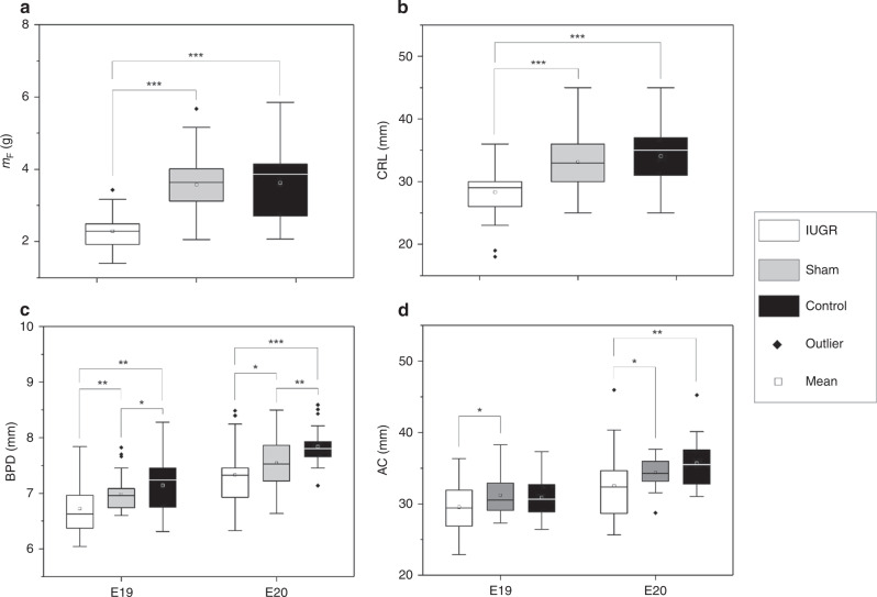
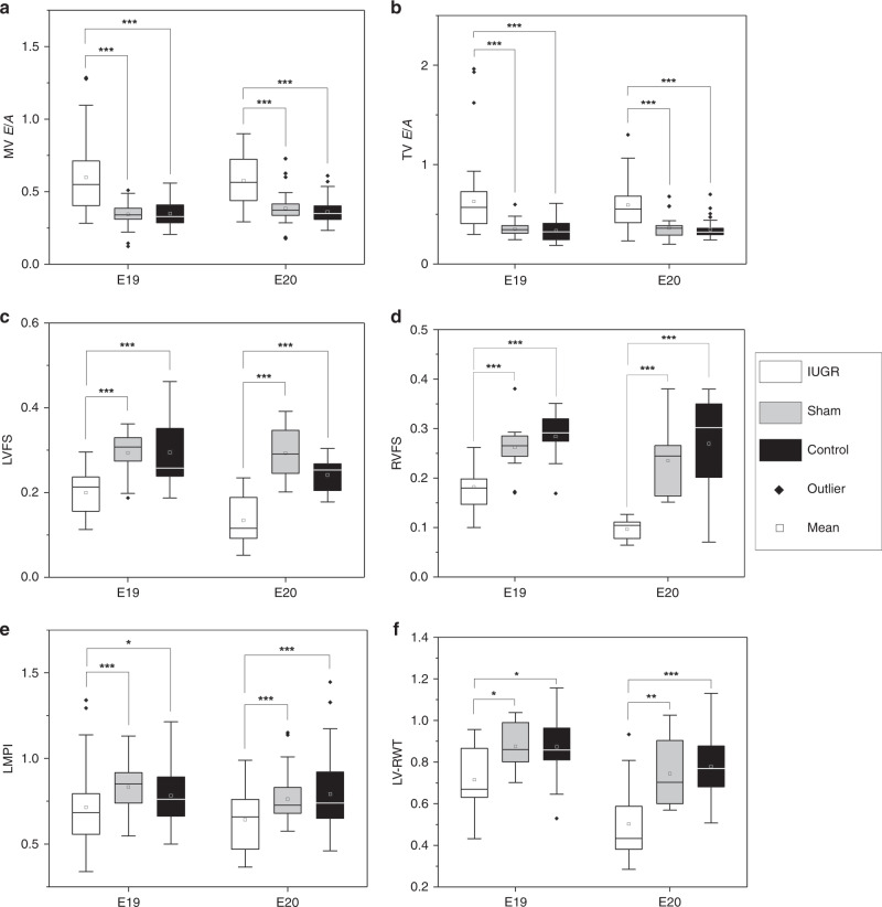
Results: Growth-restricted fetuses were significantly smaller and lighter, and had an higher placenta-to-fetus weight ratio. Growth-restricted fetal hearts had reduced wall thickness-to-diameter ratio, indicating left ventricular (LV) dilatation, and they had elevated trans-mitral and trans-tricuspid E/A ratios and reduced left and right ventricular fractional shortening (FS), suggesting systolic and diastolic dysfunction. These were similar to human IUGR fetuses. However, growth-restricted rat fetuses did not demonstrate head-sparing effect, displayed a lower LV myocardial performance index, and ventricular outflow velocities were not significantly reduced, which were dissimilar to human IUGR fetuses.
Conclusions: Despite the differences, our results suggest that this IUGR model has significant cardiac dysfunction, and could be a suitable model for studying IUGR cardiovascular physiology.
Impact: Animal models of IUGR are useful, but their fetal cardiac function is not well studied, and it is unclear if they can represent human IUGR fetuses. We performed an echocardiographic assessment of the heart function of a fetal rat model of IUGR, created via maternal uterine artery ligation. Similar to humans, the model displayed LV dilatation, elevated E/A ratios, and reduced FS. Different from humans, the model displayed reduced MPI, and no significant outflow velocity reduction. Despite differences with humans, this rat model still displayed cardiac dysfunction and is suitable for studying IUGR cardiovascular physiology.
© 2021. The Author(s).
Conflict of interest statement
The authors declare no competing interests.
Figures
References
-
- Rosenberg, A. The IUGR newborn. Semin. Perinatol.32, 219–224 (2008). - PubMed
Publication types
MeSH terms
LinkOut - more resources
Full Text Sources
Other Literature Sources
Medical
