Review
. 2021 May;22(5):346-366.
doi: 10.1038/s41580-020-00322-w. Epub 2021 Jan 27.

Diversity and versatility of p38 kinase signalling in health and disease

Affiliations
Review

Diversity and versatility of p38 kinase signalling in health and disease

Begoña Canovas et al. Nat Rev Mol Cell Biol. 2021 May.

Abstract

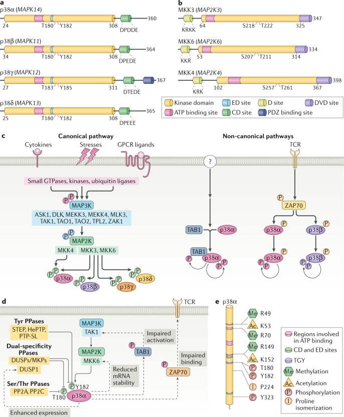
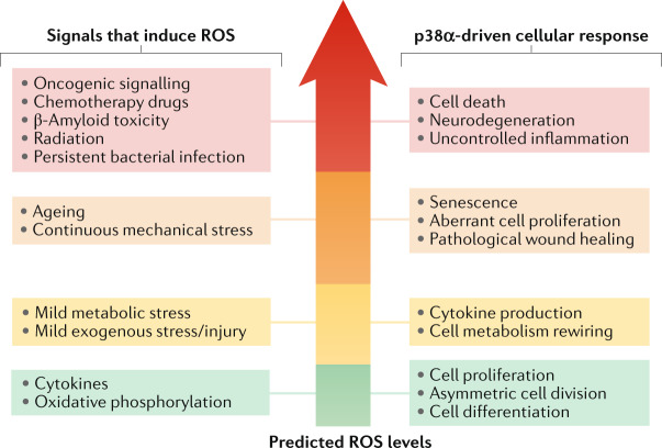
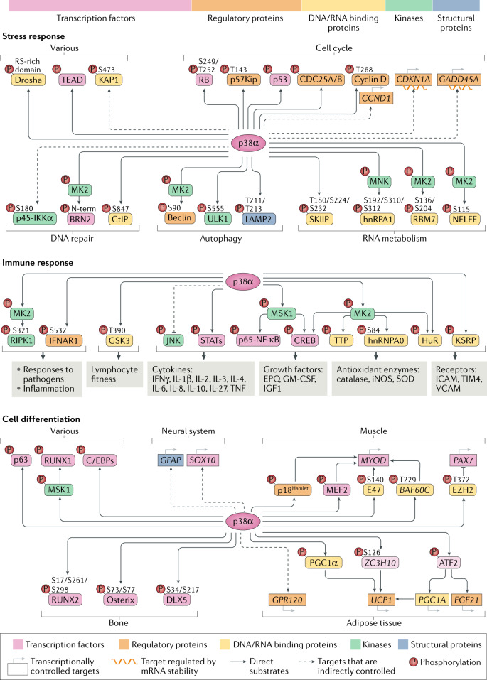
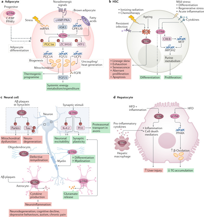
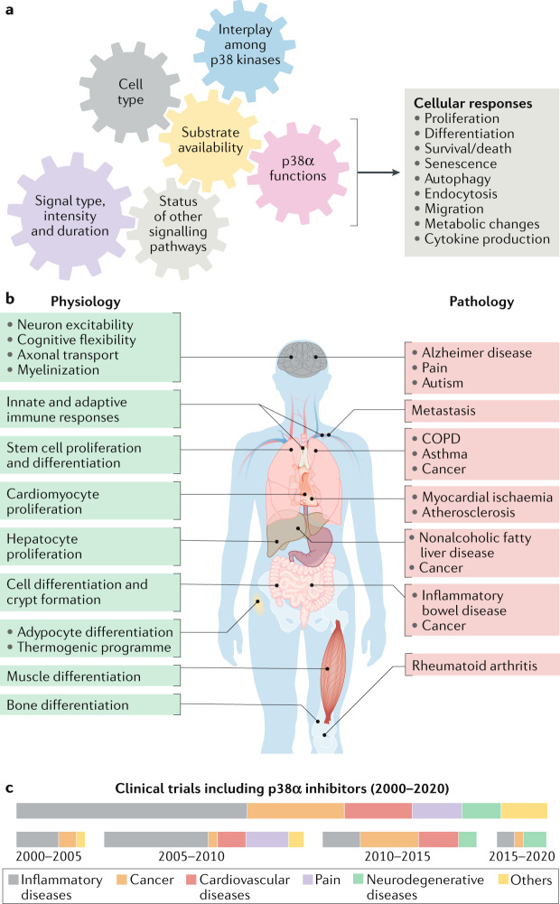
The ability of cells to deal with different types of stressful situations in a precise and coordinated manner is key for survival and involves various signalling networks. Over the past 25 years, p38 kinases - in particular, p38α - have been implicated in the cellular response to stress at many levels. These span from environmental and intracellular stresses, such as hyperosmolarity, oxidative stress or DNA damage, to physiological situations that involve important cellular changes such as differentiation. Given that p38α controls a plethora of functions, dysregulation of this pathway has been linked to diseases such as inflammation, immune disorders or cancer, suggesting the possibility that targeting p38α could be of therapeutic interest. In this Review, we discuss the organization of this signalling pathway focusing on the diversity of p38α substrates, their mechanisms and their links to particular cellular functions. We then address how the different cellular responses can be generated depending on the signal received and the cell type, and highlight the roles of this kinase in human physiology and in pathological contexts.


Conflict of interest statement

IRB Barcelona and ICREA have filed a patent application on compounds that modulate p38α autophosphorylation (WO2020120576). A.R.N. is a named inventor on this application. B.C. declares no competing interests.

Figures

Fig. 1
Fig. 1. p38 kinases and their regulation.
a | Schematic representation of the four human p38 kinases, indicating gene names (in parentheses), amino acid numbers and the different domains. The kinase domain is 90% identical in amino acid sequence among the four members. The CD domain is a negatively charged region involved in high-affinity docking interactions with substrates and regulators that contain positively charged docking (D) motifs. The ED domain contributes to substrate docking and specificity, being particularly important for interactions with mitogen-activated protein kinase (MAPK)-activated protein (MAPKAP) kinase 2 (MK2) and MK3. The ATP binding site and the phosphorylated Thr and Tyr residues of the activation loop are also indicated. p38γ has an additional carboxy-terminal region that binds to PDZ domain-containing proteins (serving as scaffolding proteins for various signalling pathways). p38 kinases are also referred to as stress-activated protein kinases: SAPK2a (p38α), SAPK2b (p38β), SAPK3 (p38γ) and SAPK4 (p38δ). b | Schematic representation of the three human MAP2Ks involved in p38 kinase activation, indicating gene names (in parentheses), amino acid numbers and highlighting the kinase domain, the ATP binding site, the D site involved in docking to p38 kinases, the DVD site that mediates interaction with MAP3Ks and the phosphorylated Ser and Thr residues of the activation loop. c | Canonical and non-canonical p38 kinase activation pathways. The colour of the phosphates (P) indicates the kinase responsible for the phosphorylation. In the canonical pathway, the first step is activation of MAP3Ks, which is triggered by various stimuli, encompassing cytokines acting via their receptors, ligands of G protein-coupled receptors (GPCRs; which include hormones, metabolites, cytokines and neurotransmitters) and stress signals. Mechanistically, MAP3Ks can be activated by multiple mechanisms, including binding to RHO, CDC45 and RAC small GTPases, phosphorylation by STE20 kinases and ubiquitylation by TRAF ubiquitin ligases, triggering phosphorylation of MAP2K, which in turn phosphorylate and activate p38 kinases. In the non-canonical pathways, activation is triggered by autophosphorylation of p38α either by binding to proteins such as transforming growth factor-β-activated protein 1 (TAB1) (observed in various cell types, but the signals responsible for activating this pathway are not well defined (question mark)) or by ZAP70 phosphorylation (specific to T cells) downstream of T cell receptor (TCR) activation. d | Scheme showing the main mechanisms leading to p38α signal termination, including phosphatases that target the activation loop phosphorylated residues, and p38α-triggered negative feedback loops (dotted lines). e | Scheme indicating human p38α protein sequence with post-translational modifications known to regulate the p38α activity. Interestingly, most modifications occur in amino acids involved in ATP binding and in the Thr-Gly-Tyr (TGY) sequence of the activation loop or near these regions. DUSP, dual-specificity phosphatase; MKP, MAPK phosphatase; PPase, protein phosphatase.
Fig. 2
Fig. 2. Interplay between ROS and p38α signalling.
Reactive oxygen species (ROS) have been reported to activate p38α in various homeostatic and pathological contexts. Importantly, ROS play essential signalling roles and their levels are known to impact cell biology in various ways. Despite the vast amount of literature linking ROS production with p38α activation, the actual levels of ROS are rarely experimentally determined. The signals reported to induce ROS and to activate p38α in different contexts are on the left, and the biological responses observed on the right. The signals and responses are organized according to the expected ROS levels in the cell, increasing from bottom to top. Lower ROS levels tend to be linked to physiological processes and homeostatic responses such as cell proliferation and differentiation or cytokine production, whereas higher ROS levels are usually generated in pathological contexts and in response to persistent stresses, eventually leading to severe cell dysfunction and death. However, how different signals trigger different ROS levels, and how diverse ROS amounts can differentially modulate p38α activation and particular biological responses remain to be fully understood.
Fig. 3
Fig. 3. The landscape of p38α substrates and targets.
p38α directly phosphorylates more than 100 proteins and can indirectly modulate a wider network of targets, explaining the versatility of this pathway. The top bar shows the relative distribution of p38α substrates according to their biological function. The panels illustrate key substrates and targets in three main p38α-regulated processes. In the stress response, p38α has been connected to many protein phosphorylation changes, which probably reflects the suitability of this mechanism for cellular adaptation by facilitating a rapid control of processes such as cell cycle progression, DNA damage repair or mRNA processing. In the immune response, p38α controls the phosphorylation of kinases, transcription factors and regulators of mRNA stability, which collectively regulate the expression of cytokines and other factors involved in inflammatory processes. In addition, the p38α pathway controls the phosphorylation of RIPK1 and the IFNα/β receptor IFNAR1, which are important in the response to pathogens and inflammation, as well as GSK3, which upon p38α phosphorylation regulates lymphocyte fitness and the adaptive immune response. In cell differentiation, and in agreement with the irreversible character of this process, p38α phosphorylates many transcription factors and chromatin modulators that will directly or indirectly control the gene expression programmes driving cell differentiation in different tissues. Dashed arrows represent indirect regulation by p38α. MK2, mitogen-activated protein kinase (MAPK)-activated protein (MAPKAP) kinase 2; RB, retinoblastoma protein; STAT, signal transducer and activator of transcription.
Fig. 4
Fig. 4. Functions of p38α in specific cell types.
The multifactorial nature of p38α signalling is illustrated by showing the diversity of functions that can be regulated by p38α depending on the cell type and the signals received. In every panel, the cell type, the extracellular stimuli (top), the signalling elements involved (when known) and the biological outcome (bottom) are indicated. Green boxes indicate homeostatic responses and red boxes pathological or deleterious events. a | p38α has a well-established role in the activation of thermogenesis in brown adipocytes. The different signals and mediators leading to p38α activation, the direct substrates and the effector targets that drive the p38α-orchestrated thermogenic programme are indicated. p38α can also regulate adipocyte differentiation, involving C/EBP and PPARγ transcription regulators of adipogenesis. Of note, contrary effects of p38α on adipogenesis have been reported depending on the model used (indicated by the split, dashed arrow), which can be linked to high dependency of p38α-mediated responses on the context as highlighted in this Review. b | The functions of p38α signalling in haematopoietic stem cells (HSCs) can be classified according to the stimuli. Upon severe or persistent stress such as infection, radiation or ageing, p38α activation correlates with elevated reactive oxygen species (ROS) levels and usually leads to detrimental responses that impair HSC function. However, in response to mild stresses, such as acute inflammation, regenerative stress or differentiation, which often involve cytokine exposure, p38α coordinates a pro-survival programme aimed to recover homeostasis. c | p38α activation has been traditionally linked to neurodegenerative diseases, especially Alzheimer disease, due to its implication in β-amyloid (Aβ) plaque formation and cytotoxicity, at least in part via its contribution to Tau hyperphosphorylation. But recent work describes additional p38α functions in different cells of the central nervous system, both in homeostasis and pathological situations. Known substrates are indicated, but targets involved in p38α-regulated synaptic plasticity, myelination or neuroinflammation remain largely unknown. There is also evidence that the role of p38α in myelination may depend on both the cause of nerve injury and the cell type. d | In hepatocytes, p38α can promote cell death or support cell viability depending on the strength of the stress: high levels of stress (such as combination of a high-fat diet (HFD) with infection/inflammation) results in cell death, whereas milder stress (such as HFD alone) generally promotes hepatocyte function in metabolizing fatty acids, by increasing their trafficking and β-oxidation, thereby reducing triglyceride (TG) storage and load in the liver. The hepatic function can be further modulated by p38α-regulated production of pro-inflammatory cytokines in macrophages that links to hepatocyte cell death.
Fig. 5
Fig. 5. Diversity of p38α roles in health and disease.
a | p38α is known to modulate many cellular processes, but not all of these functions are performed simultaneously. The gears illustrate the required coordination among several key factors that can influence the diversity of p38α-driven cellular responses. b | Genetic and pharmacological targeting of p38α in mouse models has revealed the implication of this signalling pathway in several physiological functions, and its dysregulation has been linked to a plethora of diseases. The homeostatic functions (left) and the diseases (right) in which the p38α pathway has been reported to play a role are indicated. c | Results from animal and cell-based preclinical models have supported the interest of p38α as a potential target in some of these pathologies, and several clinical trials have been developed using pharmacological p38α inhibitors, alone or in combination with other drugs. The boxes show the proportion of different pathologies targeted in clinical trials (ClinicalTrials.gov database) with p38α inhibitors (upper) and their evolution over the past 20 years (bottom). The disappointing results obtained in most clinical trials performed so far have led to a decline in the number of studies testing p38α inhibitors in patients in recent years, probably reflecting the decision of pharmaceutical companies to pursue novel targets. However, encouraging preclinical results have stimulated ongoing phase II clinical trials that either use mitogen-activated protein kinase (MAPK)-activated protein (MAPKAP) kinase 2 (MK2) inhibitors or target new diseases, as shown in Table 1. COPD, chronic obstructive pulmonary disease.

References

    1. Cuenda A, Rousseau S. p38 MAP-kinases pathway regulation, function and role in human diseases. Biochim. Biophys. Acta. 2007;1773:1358–1375. doi: 10.1016/j.bbamcr.2007.03.010. - DOI - PubMed
    1. Wagner EF, Nebreda AR. Signal integration by JNK and p38 MAPK pathways in cancer development. Nat. Rev. Cancer. 2009;9:537–549. doi: 10.1038/nrc2694. - DOI - PubMed
    1. Cuadrado A, Nebreda AR. Mechanisms and functions of p38 MAPK signalling. Biochem. J. 2010;429:403–417. doi: 10.1042/BJ20100323. - DOI - PubMed
    1. Kyriakis JM, Avruch J. Mammalian MAPK signal transduction pathways activated by stress and inflammation: a 10-year update. Physiol. Rev. 2012;92:689–737. doi: 10.1152/physrev.00028.2011. - DOI - PubMed
    1. Hotamisligil GS, Davis RJ. Cell signaling and stress responses. Cold Spring Harb. Perspect. Biol. 2016;8:a006072. doi: 10.1101/cshperspect.a006072. - DOI - PMC - PubMed

Publication types