. 2021 Feb;73(1):35-48.
doi: 10.1007/s10616-020-00440-5. Epub 2020 Nov 20.

Three-dimensional multicellular cell culture for anti-melanoma drug screening: focus on tumor microenvironment

Affiliations

Three-dimensional multicellular cell culture for anti-melanoma drug screening: focus on tumor microenvironment

Najla Adel Saleh et al. Cytotechnology. 2021 Feb.

Abstract

Abstract: The development of new treatments for malignant melanoma, which has the worst prognosis among skin neoplasms, remains a challenge. The tumor microenvironment aids tumor cells to grow and resist to chemotherapeutic treatment. One way to mimic and study the tumor microenvironment is by using three-dimensional (3D) co-culture models (spheroids). In this study, a melanoma heterospheroid model composed of cancer cells, fibroblasts, and macrophages was produced by liquid-overlay technique using the agarose gel. The size, growth, viability, morphology, cancer stem-like cells population and inflammatory profile of tumor heterospheroids and monospheroids were analyzed to evaluate the influence of stromal cells on these parameters. Furthermore, dacarbazine cytotoxicity was evaluated using spheroids and two-dimensional (2D) melanoma model. After finishing the experiments, it was observed the M2 macrophages induced an anti-inflammatory microenvironment in heterospheroids; fibroblasts cells support the formation of the extracellular matrix, and a higher percentage of melanoma CD271 was observed in this model. Additionally, melanoma spheroids responded differently to the dacarbazine than the 2D melanoma culture as a result of their cellular heterogeneity and 3D structure. The 3D model was shown to be a fast and reliable tool for drug screening, which can mimic the in vivo tumor microenvironment regarding interactions and complexity.

Keywords: 3D cell culture; Co-culture; Melanoma; Tumor microenvironment; Tumor spheroid.


Conflict of interest statement

Conflict of interestThe authors declare that they have no conflict of interest.

Figures

Fig. 1
Fig. 1
Spheroid size, viability and positive CSCs markers. a Phase contrast images of spheroids cultured at the 4th and 7th day of 3D cultivation. Scale bars represent 100 µm. b Spheroid diameter and c volume at the 4th and 7th day d cell viability and e percentage of positive CD271, CD133 and CD44 cells at the 7th day of 3D cultivation, on B and BJN spheroids (Unpaired student t-test). n = 3, each one with 8 spheroids analyzed; mean ± standard deviation. **p < 0.01, ***p < 0.001. B B16F10 monospheroids; BJN B16F10–J774–NIH/3T3 heterospheroids
Fig. 2
Fig. 2
Spheroid morphology after 7 days of 3D cultivation. a Maximum intensity projection of 20 planes of spheroids examined by confocal microscopy at 1 µm intervals stained with DAPI. Scale bars represent 100 µm b Spheroids visualized by SEM, red arrows showing a suggested presence of extracellular matrix. B B16F10 monospheroids; BJN B16F10–J774–NIH/3T3 heterospheroids
Fig. 3
Fig. 3
Spheroid histology stained with Hematoxylin & Eosin (H&E) and Mallory’s Trichrome after 7 days of 3D cultivation. The cell nuclei are stained purple by H&E and red by Mallory. The cytoplasm and extracellular matrix are stained pink by H&E and extracellular matrix is stained blue by Mallory (green arrows). Scale bars represent 100 µm. B B16F10 monospheroids; BJNB16F10–J774–NIH/3T3 heterospheroids. (Color figure online)
Fig. 4
Fig. 4
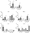
NO, IL-6, TNF-α, IL-4 and IL-10, concentrations in the supernatant of J cells, after 24 h of polarization with LPS or DEX, and spheroids prepared with J in different polarization states, after 7 days of 3D cultivation. a ·NO, b IL-6, c TNF-α, d IL-4 and e IL-10 concentrations (Unpaired student t-test). n = 3, each one with the supernatant of 8 spheroids; mean ± standard deviation; **p < 0.01; ***p < 0.001. J J774 cells; M1 J774 cells activated with lipopolysaccharides; M2 J774 cells activated with dexamethasone; B B16F10 monospheroids; BJN B16F10–J774–NIH/3T3 heterospheroids, NO nitric oxide; IL interleukin; TNF-α Tumor necrosis factor alfa. ND not detectable
Fig. 5
Fig. 5
Effect of dacarbazine after 3 days of incubation on 3D culture. a microscopy images and b graphical representations of diameter of spheroids, after 7 days of 3D cultivation, with dacarbazine Scale bar represents 100 µm (ANOVA, Bonferroni). n = 3, each one with 8 spheroids analyzed; mean ± standard deviation. *p < 0.05; **p < 0.01; ***p < 0.001. B B16F10 monospheroids; BJN B16F10–J774–NIH/3T3 heterospheroids

Similar articles

Cited by

References

    1. American Cancer Society (2019) Chemotherapy for Melanoma Skin Cancer. Available at https://www.cancer.org/cancer/melanoma-skin-cancer/treating/chemotherapy.... Accessed 6 Jun 2020
    1. Astashkina A, Mann B, Grainger DW. A critical evaluation of in vitro cell culture models for high-throughput drug screening and toxicity. Pharmacol Ther. 2012;134:82–106. doi: 10.1016/j.pharmthera.2012.01.001. - DOI - PubMed
    1. Balkwill FR, Capasso M, Hagemann T. The tumor microenvironment at a glance. J Cell Sci. 2012;125:5591–5596. doi: 10.1242/jcs.116392. - DOI - PubMed
    1. Bao B, Ahmad A, Azmi AS, et al. Cancer stem cells (CSCs) and mechanisms of their regulation: implications for cancer therapy. Curr Protoc Pharmacol. 2013 doi: 10.1002/0471141755.ph1425s61. - DOI - PMC - PubMed
    1. Bates RC, Edwards NS, Yates JD. Spheroids and cell survival. Crit Rev Oncol Hematol. 2000;36:61–74. doi: 10.1016/S1040-8428(00)00077-9. - DOI - PubMed

LinkOut - more resources