

High prevalence of hepatic steatosis and vascular thrombosis in COVID-19: A systematic review and meta-analysis of autopsy data
- PMID: 33505145
- PMCID: PMC7789052
- DOI: 10.3748/wjg.v26.i48.7693
High prevalence of hepatic steatosis and vascular thrombosis in COVID-19: A systematic review and meta-analysis of autopsy data
Abstract
Background: Coronavirus disease 2019 (COVID-19) disease can frequently affect the liver. Data on hepatic histopathological findings in COVID-19 is scarce.
Aim: To characterize hepatic pathological findings in patients with COVID-19.
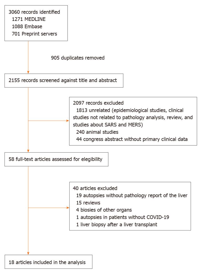
Methods: We conducted a systematic review with meta-analysis registered on PROSPERO (CRD42020192813), following PRISMA guidelines. Eligible trials were those including patients of any age and COVID-19 diagnosis based on a molecular test. Histopathological reports from deceased COVID-19 patients undergoing autopsy or liver biopsy were reviewed. Articles including less than ten patients were excluded. Proportions were pooled using random-effects models. Q statistic and I 2 were used to assess heterogeneity and levels of evidence, respectively.
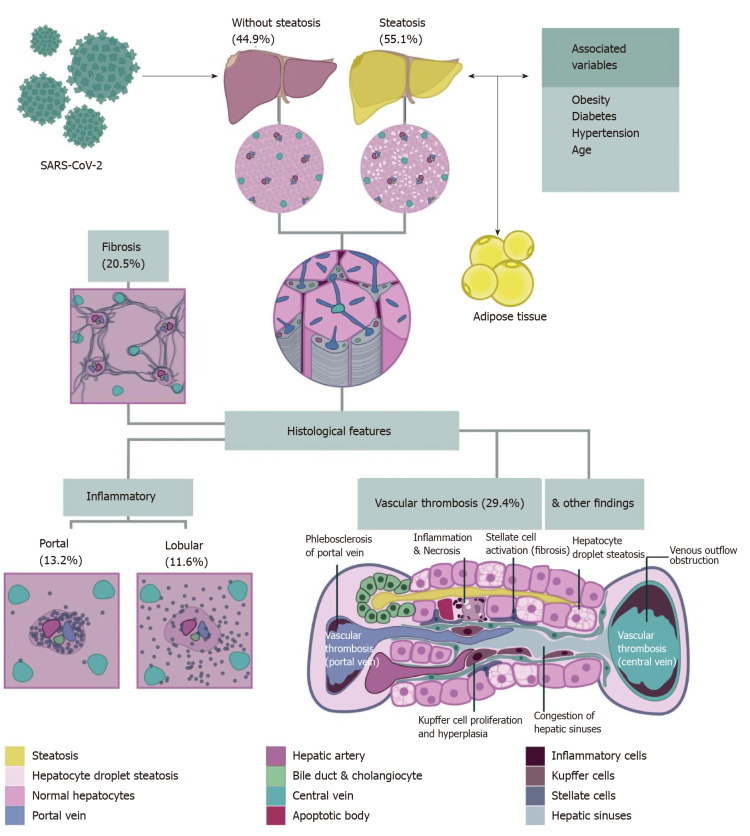
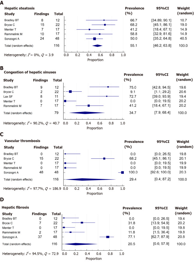
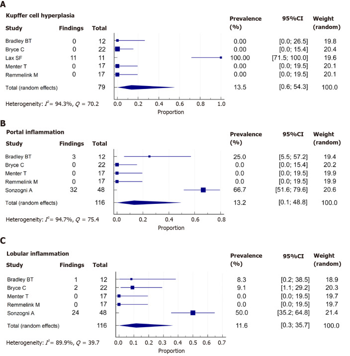
Results: We identified 18 studies from 7 countries; all were case reports and case series from autopsies. All the patients were over 15 years old, and 67.2% were male. We performed a meta-analysis of 5 studies, including 116 patients. Pooled prevalence estimates of liver histopathological findings were hepatic steatosis 55.1% [95% confidence interval (CI): 46.2-63.8], congestion of hepatic sinuses 34.7% (95%CI: 7.9-68.4), vascular thrombosis 29.4% (95%CI: 0.4-87.2), fibrosis 20.5% (95%CI: 0.6-57.9), Kupffer cell hyperplasia 13.5% (95%CI: 0.6-54.3), portal inflammation 13.2% (95%CI: 0.1-48.8), and lobular inflammation 11.6% (95%CI: 0.3-35.7). We also identified the presence of venous outflow obstruction, phlebosclerosis of the portal vein, herniated portal vein, periportal abnormal vessels, hemophagocytosis, and necrosis.
Conclusion: We found a high prevalence of hepatic steatosis and vascular thrombosis as major histological liver features. Other frequent findings included portal and lobular inflammation and Kupffer cell hyperplasia or proliferation. Further studies are needed to establish the mechanisms and implications of these findings.
Keywords: Autopsies; COVID-19; Liver; Liver biopsies; Pathology; SARS-CoV-2.
©The Author(s) 2020. Published by Baishideng Publishing Group Inc. All rights reserved.
Conflict of interest statement
Conflict-of-interest statement: The authors have nothing to disclose.
Figures
References
-
- Cheung KS, Hung IFN, Chan PPY, Lung KC, Tso E, Liu R, Ng YY, Chu MY, Chung TWH, Tam AR, Yip CCY, Leung KH, Fung AY, Zhang RR, Lin Y, Cheng HM, Zhang AJX, To KKW, Chan KH, Yuen KY, Leung WK. Gastrointestinal Manifestations of SARS-CoV-2 Infection and Virus Load in Fecal Samples From a Hong Kong Cohort: Systematic Review and Meta-analysis. Gastroenterology. 2020;159:81–95. - PMC - PubMed
-
- Sultan S, Altayar O, Siddique SM, Davitkov P, Feuerstein JD, Lim JK, Falck-Ytter Y, El-Serag HB AGA Institute. AGA Institute Rapid Review of the Gastrointestinal and Liver Manifestations of COVID-19, Meta-Analysis of International Data, and Recommendations for the Consultative Management of Patients with COVID-19. Gastroenterology 2020; 159: 320-334. :e27. - PMC - PubMed
Publication types
MeSH terms
LinkOut - more resources
Full Text Sources
Medical
Miscellaneous
