Review
. 2021 Jan 14:2021:8874339.
doi: 10.1155/2021/8874339. eCollection 2021.

COVID-19 and Toll-Like Receptor 4 (TLR4): SARS-CoV-2 May Bind and Activate TLR4 to Increase ACE2 Expression, Facilitating Entry and Causing Hyperinflammation

Affiliations
Review

COVID-19 and Toll-Like Receptor 4 (TLR4): SARS-CoV-2 May Bind and Activate TLR4 to Increase ACE2 Expression, Facilitating Entry and Causing Hyperinflammation

Mohamed M Aboudounya et al. Mediators Inflamm. .

Abstract

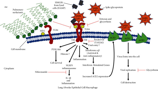
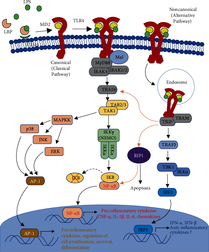
Causes of mortality from COVID-19 include respiratory failure, heart failure, and sepsis/multiorgan failure. TLR4 is an innate immune receptor on the cell surface that recognizes pathogen-associated molecular patterns (PAMPs) including viral proteins and triggers the production of type I interferons and proinflammatory cytokines to combat infection. It is expressed on both immune cells and tissue-resident cells. ACE2, the reported entry receptor for SARS-CoV-2, is only present on ~1-2% of the cells in the lungs or has a low pulmonary expression, and recently, the spike protein has been proposed to have the strongest protein-protein interaction with TLR4. Here, we review and connect evidence for SARS-CoV-1 and SARS-CoV-2 having direct and indirect binding to TLR4, together with other viral precedents, which when combined shed light on the COVID-19 pathophysiological puzzle. We propose a model in which the SARS-CoV-2 spike glycoprotein binds TLR4 and activates TLR4 signalling to increase cell surface expression of ACE2 facilitating entry. SARS-CoV-2 also destroys the type II alveolar cells that secrete pulmonary surfactants, which normally decrease the air/tissue surface tension and block TLR4 in the lungs thus promoting ARDS and inflammation. Furthermore, SARS-CoV-2-induced myocarditis and multiple-organ injury may be due to TLR4 activation, aberrant TLR4 signalling, and hyperinflammation in COVID-19 patients. Therefore, TLR4 contributes significantly to the pathogenesis of SARS-CoV-2, and its overactivation causes a prolonged or excessive innate immune response. TLR4 appears to be a promising therapeutic target in COVID-19, and since TLR4 antagonists have been previously trialled in sepsis and in other antiviral contexts, we propose the clinical trial testing of TLR4 antagonists in the treatment of severe COVID-19. Also, ongoing clinical trials of pulmonary surfactants in COVID-19 hold promise since they also block TLR4.


Conflict of interest statement

The authors declare that they have no conflict of interest.

Figures

Figure 1
Figure 1
Graphical abstract. Note that less pulmonary surfactant secretion leads to exposed TLR4 receptors on alveolar cells. HMGB1 released from lysed cells can also activate TLR4 causing excessive inflammation and fibrosis. The same model applies to a cardiac cell, except that there would be no pulmonary surfactants, and instead of the outside air, it would be the extracellular matrix. In the heart, TLR4 activation by the SARS-CoV-2 virus and/or DAMPs released from infected, necrotic cells or even upregulated at sites of injury may also cause abnormal signalling towards the canonical proinflammatory pathway rather than the alternative anti-inflammatory pathway. This would cause viral myocarditis. TLR4 activation also decreases the contractility of cardiomyocytes. In addition, SARS-CoV-2 may activate TLR4 to increase PI3K/Akt signalling in infected cells, preventing apoptosis and thus increasing time for viral replication. Aberrant inflammatory signalling could also be extended to other tissues expressing TLR4, such as the skin and kidney, where the virus would therefore cause multiple-organ injury.
Figure 2
Figure 2
Overview of the main TLR4 signalling pathways. TLR4 can be activated by LPS (classical PAMP), DAMPs, or viral PAMPs. LPS is also picked up by the LPS-binding protein (LBP) present in the blood and in extracellular fluid in tissues. LPS is transferred from LBP to the coreceptor CD14. Recognition by MD2 and TLR4 binding results in the nuclear induction of transcription factors NF-κB and AP-1 via the MyD88-dependent pathway involving IRAKs, TABs, TAK, MAP kinases, and IKK isoforms. This results in the transcription of proinflammatory cytokines and regulators of cell proliferation, survival, and differentiation. In fibroblasts, this pathway mediates myofibroblast differentiation and results in CTGF and collagen production [100], while the alternative pathway is endosomal and involves TRIF and TRAM complex formation at the TIR domain. The alternative pathway results in the expression of anti-inflammatory cytokines, interferons α and β regulated by interferon regulatory factor 3 (IRF3), in addition to some NF-κB nuclear translocation. “Cross-talk” activation between the two pathways is indicated by brown arrows. Adaptors and cascade proteins are usually in homodimers, like the TLR4 receptor [22], but were omitted here for simplicity.

Similar articles

Cited by

References

    1. World health organization. Coronavirus disease (COVID-2019) situation reports. 2020. https://www.who.int/emergencies/diseases/novel-coronavirus-2019/situatio...
    1. Huang C., Wang Y., Li X., et al. Clinical features of patients infected with 2019 novel coronavirus in Wuhan, China. The lancet. 2020;395(10223):497–506. doi: 10.1016/S0140-6736(20)30183-5. - DOI - PMC - PubMed
    1. Guan W. J., Ni Z. Y., Hu Y., et al. Clinical characteristics of coronavirus disease 2019 in China. The New England Journal of Medicine. 2020;382(18):1708–1720. doi: 10.1056/NEJMoa2002032. - DOI - PMC - PubMed
    1. Tufan A., Avanoglu Guler A., Matucci-Cerinic M. COVID-19, immune system response, hyperinflammation and repurposing antirheumatic drugs. Turkish Journal Of Medical Sciences. 2020;50(SI-1):620–632. doi: 10.3906/sag-2004-168. - DOI - PMC - PubMed
    1. Shereen M. A., Khan S., Kazmi A., Bashir N., Siddique R. COVID-19 infection: origin, transmission, and characteristics of human coronaviruses. Journal of Advanced Research. 2020;24:91–98. doi: 10.1016/j.jare.2020.03.005. - DOI - PMC - PubMed