

Zisheng Shenqi Decoction Ameliorates Monosodium Urate-Mediated Gouty Arthritis in Rats via Promotion of Autophagy through the AMPK/mTOR Signaling Pathway
- PMID: 33505502
- PMCID: PMC7806400
- DOI: 10.1155/2021/6918026
Zisheng Shenqi Decoction Ameliorates Monosodium Urate-Mediated Gouty Arthritis in Rats via Promotion of Autophagy through the AMPK/mTOR Signaling Pathway
Abstract
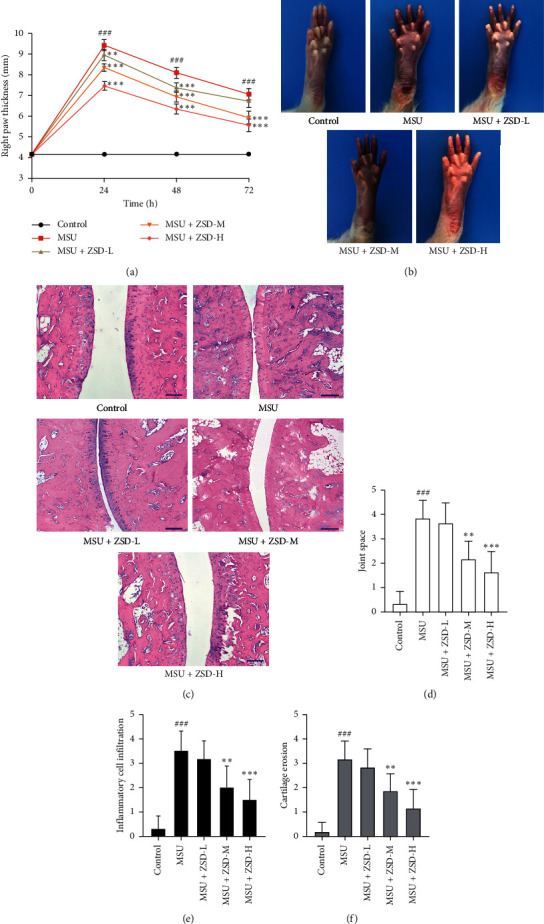
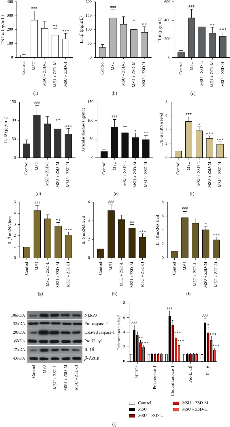
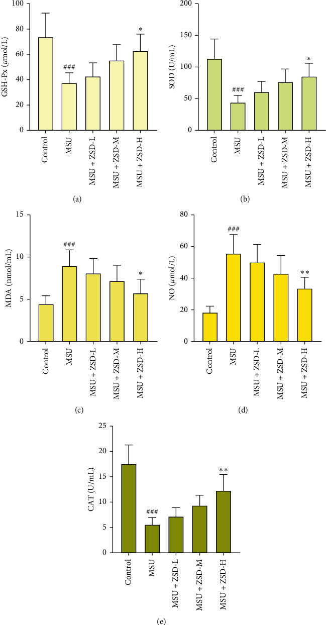
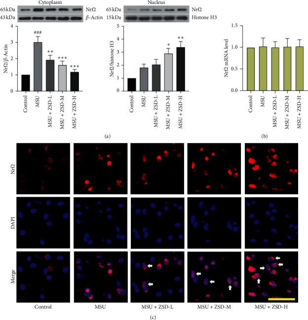
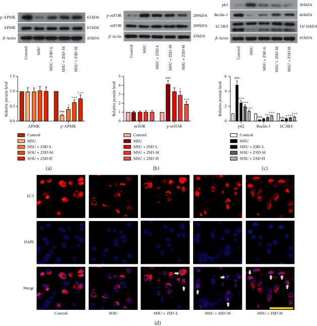
Gouty arthritis (GA) is an inflammatory disease owing to the accumulation of monosodium urate (MSU) in joints, leading to redness and burning pain. In this study, the effect of Zisheng Shenqi Decoction (ZSD) on a rat model of MSU-induced GA was investigated. ZSD obviously diminished the right paw thickness, the degree of the swelling of the paw, and the infiltration of the inflammatory cell, as well as cartilage erosion, and widened the joint space in MSU-treated rats. Besides, MSU remarkably elevated the release of tumor necrosis factor-α (TNF-α), interleukin-1β (IL-1β), IL-6, and IL-18; however, ZSD treatment dose dependently lowered these levels and resulted in a significant decrease in articular elastase activity. Also, ZSD administration increased the activities of superoxide dismutase (SOD), glutathione peroxidase (GSH-Px), and catalase (CAT) but declined malondialdehyde (MDA) and nitrogen monoxide (NO) contents. Importantly, western blotting analysis revealed that NOD-like receptor protein 3 (NLRP3), cleaved caspase-1, IL-1β, nuclear factor-E2-related factor 2 (Nrf2) in the cytoplasm, phosphorylated mammalian target of rapamyclin (p-mTOR), and p62 expressions were downregulated, whereas the levels of nuclear Nrf2, phosphorylated AMP-activated protein kinase (p-AMPK), Beclin-1, and LC3II/I were upregulated by ZSD. Immunofluorescence assay indicated that ZSD evidently promoted nuclear translocation of LC3. Taken together, ZSD inhibited inflammation and oxidative stress and facilitated autophagy through the activation of the AMPK pathway and suppression of the mTOR signaling pathway, demonstrating its potential for preventing and curing GA.
Copyright © 2021 Jieru Han et al.
Conflict of interest statement
The authors declare that they have no conflicts of interest.
Figures
Similar articles
-
Zisheng Shenqi decoction ameliorates monosodium urate crystal-induced gouty arthritis in rats through anti-inflammatory and anti-oxidative effects.Mol Med Rep. 2016 Sep;14(3):2589-97. doi: 10.3892/mmr.2016.5526. Epub 2016 Jul 18. Mol Med Rep. 2016. PMID: 27432278 Free PMC article.
-
Exploration of the mechanism of Zisheng Shenqi decoction against gout arthritis using network pharmacology.Comput Biol Chem. 2021 Feb;90:107358. doi: 10.1016/j.compbiolchem.2020.107358. Epub 2020 Aug 8. Comput Biol Chem. 2021. PMID: 33243703 Review.
-
Curcumin ameliorates monosodium urate-induced gouty arthritis through Nod-like receptor 3 inflammasome mediation via inhibiting nuclear factor-kappa B signaling.J Cell Biochem. 2019 Apr;120(4):6718-6728. doi: 10.1002/jcb.27969. Epub 2018 Dec 28. J Cell Biochem. 2019. PMID: 30592318
-
Berberine, an isoquinoline alkaloid suppresses TXNIP mediated NLRP3 inflammasome activation in MSU crystal stimulated RAW 264.7 macrophages through the upregulation of Nrf2 transcription factor and alleviates MSU crystal induced inflammation in rats.Int Immunopharmacol. 2017 Mar;44:26-37. doi: 10.1016/j.intimp.2016.12.031. Epub 2017 Jan 7. Int Immunopharmacol. 2017. PMID: 28068647
-
Morin, a dietary bioflavonol suppresses monosodium urate crystal-induced inflammation in an animal model of acute gouty arthritis with reference to NLRP3 inflammasome, hypo-xanthine phospho-ribosyl transferase, and inflammatory mediators.Eur J Pharmacol. 2016 Sep 5;786:116-127. doi: 10.1016/j.ejphar.2016.06.005. Epub 2016 Jun 3. Eur J Pharmacol. 2016. PMID: 27268719
Cited by
-
Natural Xanthine Oxidase Inhibitor 5-O-Caffeoylshikimic Acid Ameliorates Kidney Injury Caused by Hyperuricemia in Mice.Molecules. 2021 Dec 1;26(23):7307. doi: 10.3390/molecules26237307. Molecules. 2021. PMID: 34885887 Free PMC article.
-
Daphnetin alleviates inflammation and promotes autophagy via the AMPK/mTOR pathway in gouty arthritis.J Cell Commun Signal. 2025 Apr 28;19(2):e70011. doi: 10.1002/ccs3.70011. eCollection 2025 Jun. J Cell Commun Signal. 2025. PMID: 40304008 Free PMC article.
-
HDAC8 Promotes Liver Metastasis of Colorectal Cancer via Inhibition of IRF1 and Upregulation of SUCNR1.Oxid Med Cell Longev. 2022 Aug 16;2022:2815187. doi: 10.1155/2022/2815187. eCollection 2022. Oxid Med Cell Longev. 2022. PMID: 36035205 Free PMC article.
-
Zhuanggu Zhitong Capsule alleviates postmenopausal osteoporosis in ovariectomized rats by regulating autophagy through AMPK/mTOR signaling pathway.Ann Transl Med. 2022 Aug;10(16):900. doi: 10.21037/atm-22-3724. Ann Transl Med. 2022. PMID: 36111039 Free PMC article.
-
Medicinal fungus Phellinus igniarius alleviates gout in vitro by modulating TLR4/NF-kB/NLRP3 signaling.Front Pharmacol. 2022 Oct 21;13:1011406. doi: 10.3389/fphar.2022.1011406. eCollection 2022. Front Pharmacol. 2022. PMID: 36339594 Free PMC article.
References
LinkOut - more resources
Full Text Sources
Other Literature Sources
Miscellaneous
