[Confusion in the diagnosis of endobronchial tumor]
- PMID: 33505570
- PMCID: PMC7813657
- DOI: 10.11604/pamj.2020.37.201.22896
[Confusion in the diagnosis of endobronchial tumor]
Abstract
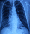
Bronchopulmonary cancer is the leading cause of death in men and the second in women. Some endoscopic or radiological features may guide histological diagnosis and thus facilitate therapeutic management. We here report the case of a 54-year old man, with a history of smoking and recent coronary stent implantation, presenting with haemoptysis and worsening of dyspnea which had evolved over the last month. Chest x-ray showed left pulmonary hemifield lucency with signs of retraction. Bronchial fibroscopy objectified raspberry bud formation spontaneously bleeding, originating from the left main bronchus and suggesting carcinoid tumor. Chest computed tomography (CT) scan showed poorly enhanced endoluminal tissue process at the level of the left main bronchus, located four cm from the carina and complicated with atelectasis. Diagnostic and therapeutic surgery helped to adjust to a diagnosis of endobronchial amartocondroma.
Le cancer broncho-pulmonaire représente la première cause de décès par cancer chez l´homme et le deuxième chez la femme. Certaines présentations endoscopiques ou radiologiques peuvent orienter le diagnostic histologique et ainsi faciliter la prise en charge thérapeutique. Nous rapportons l´observation d´un homme de 54 ans, tabagique, coronarien récemment stenté, consultant pour hémoptysie et aggravation de sa dyspnée évoluant depuis un mois. Sa radiographie du thorax avait objectivé une hyperclarté de l´hémichamp pulmonaire gauche avec des signes de rétraction. A la fibroscopie bronchique, il existait une formation bourgeonnante framboisée, saignant spontanément, accouchée par la bronche souche gauche évoquant une tumeur carcinoïde. Le scanner thoracique avait objectivé un processus tissulaire endoluminal, au niveau de la bronche souche gauche situé à quatre cm de la carène, peu rehaussé au produit de contraste et compliqué d´atélectasie. Une chirurgie diagnostique et thérapeutique a permis de redresser le diagnostic en faveur d´un hamartochondrome endobronchique.
Keywords: Bronchial fibroscopy; endobronchial tumor; thoracic CT scan.
Copyright: Houda Snène et al.
Conflict of interest statement
Les auteurs ne déclarent aucun conflit d´intérêts.
Figures


References
-
- Shah H, Garbe L, Nussbaum E, Dumon JF, Chiodera PL, Cavaliere S. Benign tumors of the tracheobronchial tree: endoscopic characteristics and role of laser resection. Chest. 1995 Jun;107(6):1744–51. - PubMed
-
- Travis WD, Brambilla E, Müller-Hermelink H, Harris CC. Pathology and genetics of tumours of the Lung, pleura, thymus and heart. Lyon: IARC Press; 2004. World Health Organisation classification of tumours.
-
- Godwin JD. Carcinoid tumors, An analysis of 2.837 cases. Cancer. 1975 Aug;36(2):560–569. - PubMed
-
- Gjevre JA, Myers JL, Prakash UB. Pulmonary hamartomas. Mayo Clin Proc. 1996 Jan;71(1):14–20. - PubMed
-
- Cosío BG, Villena V, Echave-Sustaeta J, De Miguel E, Alfaro J, Hernandez L, et al. Endobronchial Hamartoma. Chest. 2002 Jul;122(1):202–5. - PubMed
Publication types
MeSH terms
LinkOut - more resources
Full Text Sources
Medical
