

Evaluation of microleakage in class-II bulk-fill composite restorations
- PMID: 33505621
- PMCID: PMC7816009
- DOI: 10.1016/j.jds.2020.04.007
Evaluation of microleakage in class-II bulk-fill composite restorations
Abstract
Background/purpose: Despite the clinical appeal of restoring deep class II cavities in single increment using bulk-fill resin composite, sealing of bulk-filled composite restorations is a concern. This study evaluated interfacial adaptation of bulk-fill composite restoration to axial wall and gingival floor of class II cavities using cross-polarization optical coherence tomography (CP-OCT).
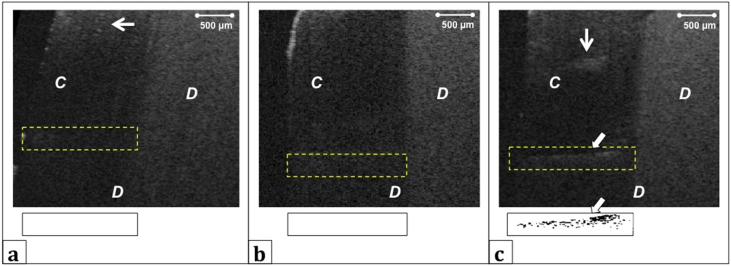
Materials and methods: Box-shaped class II cavities were prepared in extracted molars and divided into three groups (n = 7) according to adhesive used; Clearfil SE Bond 2 (SE2), Tetric-N Bond Self-Etch (TSE) or Tetric-N Bond Universal (TNU). All adhesives were applied in self-etch mode and according to manufacturers' recommendation. Then, preparations were bulk-filled with Filtek Bulk Fill Posterior Restorative resin composite and immersed in a contrast agent. Tomographic images of axial wall and gingival floor of each restoration were obtained by CP-OCT (IVS-300, Santec) with a central wavelength of 1330 nm and were imported to an image analysis software to quantify microleakage.
Results: Mann-Whitney U test showed statistically significant difference in microleakage percentage between the groups at both axial wall and gingival floor (p < 0.05). SE2 group had the lowest percentage of microleakage (p < 0.05), as only few cross-sections showed areas of reflections from contrast agent penetrating into axial wall (8.23 ± 6.8) and gingival floor (7.07 ± 4.1), followed by TNU group (18.13 ± 12.9 axially and 30.61 ± 11.9 gingivally). Microleakage was frequently observed at the axial wall and gingival floor of TSE group, showing the highest percentages of 25.50 ± 12.5 and 36.97 ± 10.2, respectively (p < 0.05).
Conclusion: All tested groups exhibited different extent of interfacial microleakage, however, two-step self-etch adhesive yielded superior adaptation in comparison to one-step self-etch adhesive and universal adhesive.
Keywords: Adhesive; Bulk-filled; Composite-resins; Microleakage; Optical coherence tomography.
© 2020 Association for Dental Sciences of the Republic of China. Publishing services by Elsevier B.V.
Conflict of interest statement
No competing financial interests exist.
Figures


References
-
- Leprince J.G., Palin W.M., Vanacker J., Sabbagh J., Devaux J., Leloup G. Physico-mechanical characteristics of commercially available bulk-fill composites. J Dent. 2014;42:993–1000. - PubMed
-
- Ferracane J.L. Resin composite -- state of the art. Dent Mater. 2011;27:29–38. - PubMed
-
- Ferracane J.L., Hilton T.J. Polymerization stress -- is it clinically meaningful? Dent Mater. 2016;32:1–10. - PubMed
-
- Li X., Pongprueksa P., Van Meerbeek B., De Munck J. Curing profile of bulk-fill resin-based composites. J Dent. 2015;43:664–672. - PubMed
-
- Bucuta S., Ilie N. Light transmittance and micro-mechanical properties of bulk fill vs. conventional resin based composites. Clin Oral Investig. 2014;18:1991–2000. - PubMed
LinkOut - more resources
Full Text Sources
Miscellaneous
