

Evaluation of Mental Foramen with Cone Beam Computed Tomography: A Systematic Review of Literature
- PMID: 33505723
- PMCID: PMC7806401
- DOI: 10.1155/2021/8897275
Evaluation of Mental Foramen with Cone Beam Computed Tomography: A Systematic Review of Literature
Abstract
Purpose: The aim of this systematic review is to assess whether the anatomy of mental foramen is precisely evaluable with cone beam computed tomography (CBCT) before implantation in humans.
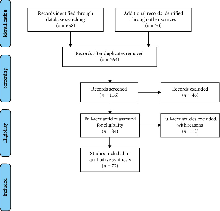
Methods: A systematic review was carried out to evaluate the anatomy of mental foramen (size, position, symmetry, anterior loop, and accessory mental foramen or multiple mental foramina). According to Preferred Reporting Items for Systematic reviews and Meta-Analyses (PRISMA) guidelines, an electronic search of three databases (Medline, Web of Science, and Cochrane Library) was undertaken until June 2020 and was supplemented by manual searching. Two reviewers will independently perform the processes of study inclusion, data extraction, and quality assessment. Systematic reviews, studies about children, and case reports were excluded. Only studies using CBCT to do preoperative evaluation were selected.
Results: From 728 potentially eligible articles, 72 were included in the qualitative analysis and quantitative synthesis. This systematic review provided an assessment of the anatomy of the mental foramen. The mental foramen was located mostly between the two premolars (between 50.4% and 61.95%) or apically to the second premolar (from 50.3% to 57.9%). The mean diameter of the mental foramen was bigger in males than in females; the difference between them could reach 0.62 mm. The anterior loop seemed to be longer in males (between 0.87 ± 1.81 and 7.25 ± 2.02 mm) than in females (between 0.81 ± 1.18 and 6.52 ± 1.63 mm) and with the presence of teeth (from 0.91 ± 1.18 to 2.55 ± 1.28 for dentate people and from 0.25 ± 0.61 to 2.40 ± 0.88 mm for edentate population). The anterior loop and the accessory mental foramina were detected more frequently with CBCT than panoramic X-ray: only between 0.0 and 48.6% AMFs detected with CBCT were also seen with panoramic images. Clinical Significance. The mental foramen (MF) is an important landmark for local anesthesia and surgical and implantology procedures. Its location, morphology, and anatomical variations need to be considered to avoid mental nerve injury. The aim of this review is to evaluate the mental foramen using CBCT through a systematic literature review to improve knowledge of this complex area for the clinician.
Copyright © 2021 Antoinette Pelé et al.
Conflict of interest statement
The authors declare that they have no conflicts of interest.
Figures

Similar articles
-
Evaluation of mental foramen and accessory mental foramen using cone beam computed tomography in a Turkish population.BMC Med Imaging. 2025 Apr 30;25(1):140. doi: 10.1186/s12880-025-01589-1. BMC Med Imaging. 2025. PMID: 40307719 Free PMC article.
-
Morphometric Analysis of the Mandibular Canal, Anterior Loop, and Mental Foramen: A Cone-Beam Computed Tomography Evaluation.Int J Environ Res Public Health. 2021 Mar 24;18(7):3365. doi: 10.3390/ijerph18073365. Int J Environ Res Public Health. 2021. PMID: 33805123 Free PMC article.
-
A Limited Field Cone-beam Computed Tomography-based Evaluation of the Mental Foramen, Accessory Mental Foramina, Anterior Loop, Lateral Lingual Foramen, and Lateral Lingual Canal.J Endod. 2018 Jun;44(6):946-951. doi: 10.1016/j.joen.2018.01.013. Epub 2018 Mar 15. J Endod. 2018. PMID: 29550007
-
Identification of anterior loop in different populations to avoid nerve injury during surgical procedures-a systematic review and meta-analysis.Oral Maxillofac Surg. 2021 Jun;25(2):159-174. doi: 10.1007/s10006-020-00915-x. Epub 2020 Oct 29. Oral Maxillofac Surg. 2021. PMID: 33118108
-
The mental foramen and nerve: clinical and anatomical factors related to dental implant placement: a literature review.J Periodontol. 2006 Dec;77(12):1933-43. doi: 10.1902/jop.2006.060197. J Periodontol. 2006. PMID: 17209776 Review.
Cited by
-
Morphological Characteristics of the Double Mental Foramen and Its Relevance in Clinical Practice: An Observational Study.Diagnostics (Basel). 2024 Jun 17;14(12):1277. doi: 10.3390/diagnostics14121277. Diagnostics (Basel). 2024. PMID: 38928695 Free PMC article.
-
Anatomical characteristics of mental foramen and canal: A cone-beam computed tomography analysis.J Clin Exp Dent. 2024 Aug 1;16(8):e1004-e1011. doi: 10.4317/jced.61861. eCollection 2024 Aug. J Clin Exp Dent. 2024. PMID: 39281795 Free PMC article.
-
Accessory lingual mental foramen: A case report of a rare anatomic variation.Oral Radiol. 2024 Jul;40(3):410-414. doi: 10.1007/s11282-024-00747-5. Epub 2024 Mar 25. Oral Radiol. 2024. PMID: 38523181
-
Evaluating the Anterior Loop of the Mental Nerve Using Cone Beam CT Scans in the Jordanian Population.Cureus. 2024 Apr 18;16(4):e58519. doi: 10.7759/cureus.58519. eCollection 2024 Apr. Cureus. 2024. PMID: 38957815 Free PMC article.
-
Anatomical variant of anteriorly extending mental foramen: A case report.Clin Case Rep. 2024 Jan 4;12(1):e8341. doi: 10.1002/ccr3.8341. eCollection 2024 Jan. Clin Case Rep. 2024. PMID: 38188843 Free PMC article.
References
-
- Solar P., Ulm C., Frey G., Matejka M. A classification of the intraosseous paths of the mental nerve. International Journal of Oral Maxillofacial Implants. 1994;9:339–344.
-
- Demir A., Izgi E., Pekiner F. Anterior loop of the mental foramen in a Turkish subpopulation with dentate patients: a cone beam computed tomography study. Müsbed. 2015;5:231–238.
Publication types
LinkOut - more resources
Full Text Sources
Other Literature Sources
