

Kimura's Disease: A Rare Cause of Unilateral Tonsillar Enlargement
- PMID: 33505749
- PMCID: PMC7808821
- DOI: 10.1155/2021/8815317
Kimura's Disease: A Rare Cause of Unilateral Tonsillar Enlargement
Abstract
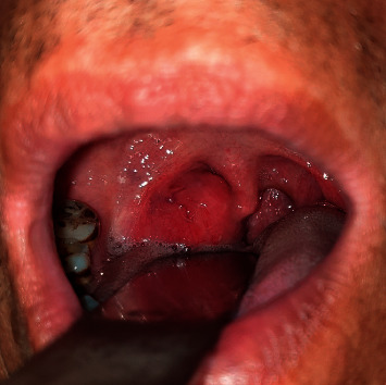
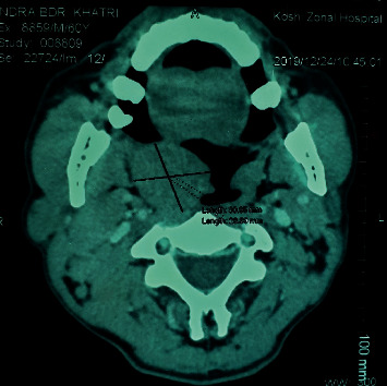
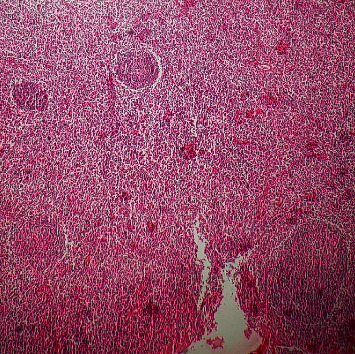
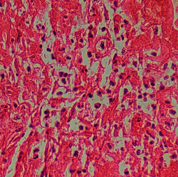
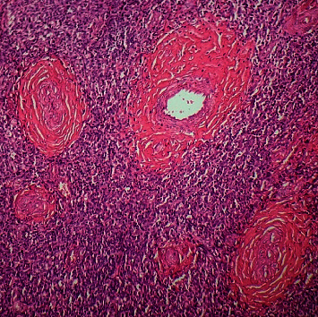
Introduction: Kimura's disease is a rare inflammatory disorder of unknown cause, commonly seen in young Asian males. Case Report. A 61-year-old male patient presented with a history of right tonsillar mass and cervical lymphadenopathy. The patient underwent hematological investigation and imaging followed by resection of tonsillar mass. Based on histopathological and subsequent immunohistochemistry reports, the case was diagnosed as Kimura's disease of the tonsil. Discussion. Kimura's disease commonly presents as painless subcutaneous masses in the head and neck region or cervical lymphadenopathy. Kimura's disease presenting as a tonsillar mass is a very rare condition. Patients usually have peripheral eosinophilia and elevated levels of serum IgE. The diagnosis is based on the clinical and histopathologic findings in a biopsy of the mass and/or lymph node along with elevated peripheral eosinophil and serum IgE level.
Conclusion: The clinical presentation of Kimura's disease is highly variable. Kimura's disease should be considered as a differential diagnosis in patients presenting with a tonsillar mass. A high index of suspicion along with histopathological examination helps in the early diagnosis and management. Surgical excision is the treatment of choice.
Copyright © 2021 Prakash Khanal and Agya Shrestha.
Conflict of interest statement
The authors declare that there are no conflicts of interest regarding the publication of this paper.
Figures
Similar articles
-
Kimura's disease - An unusual presentation involving subcutaneous tissue, parotid gland and lymph node.J Oral Maxillofac Pathol. 2013 Sep;17(3):455-9. doi: 10.4103/0973-029X.125220. J Oral Maxillofac Pathol. 2013. PMID: 24574673 Free PMC article.
-
Eosinophilic peritonitis and nephrotic syndrome in Kimura's disease: a case report and literature review : Eosinophilic peritonitis in Kimura's disease.BMC Nephrol. 2020 Apr 17;21(1):138. doi: 10.1186/s12882-020-01791-z. BMC Nephrol. 2020. PMID: 32303193 Free PMC article.
-
Kimura's disease: A diagnostic challenge experienced with cytology of postauricular swelling with histopathological relevance.J Cytol. 2016 Oct-Dec;33(4):232-235. doi: 10.4103/0970-9371.190453. J Cytol. 2016. PMID: 28028342 Free PMC article.
-
Kimura's disease successively affecting multiple body parts: a case-based literature review.BMC Ophthalmol. 2022 Apr 2;22(1):154. doi: 10.1186/s12886-022-02378-y. BMC Ophthalmol. 2022. PMID: 35366827 Free PMC article. Review.
-
Kimura's disease sequentially involving multiple sites in the head and neck: A case report with a 13-year follow-up and literature review.J Int Med Res. 2025 May;53(5):3000605251337422. doi: 10.1177/03000605251337422. Epub 2025 May 13. J Int Med Res. 2025. PMID: 40357910 Free PMC article. Review.
Cited by
-
A 23-year-old male patient with Kimura's disease without renal transplantation: a rare case report from Syria.Ann Med Surg (Lond). 2024 Jul 1;86(8):4927-4931. doi: 10.1097/MS9.0000000000002341. eCollection 2024 Aug. Ann Med Surg (Lond). 2024. PMID: 39118751 Free PMC article.
References
-
- Ioachim H., Ratech H. Kimura lymphadenopathy. In: Ioachim H., Ratech H., editors. Ioachim’s Lymph Node Pathology. 3rd. Philadelphia, PA, USA: Lippincott-Raven; 2002. pp. 209–211.
Publication types
LinkOut - more resources
Full Text Sources
Other Literature Sources
