Case Reports
. 2020 Dec 23;12(12):e12232.
doi: 10.7759/cureus.12232.

Tenosynovial Giant Cell Tumor of the Cervical Spine: Case Report and Review of the Literature

Affiliations
Case Reports

Tenosynovial Giant Cell Tumor of the Cervical Spine: Case Report and Review of the Literature

Meena Thatikunta et al. Cureus. .

Abstract

Tenosynovial giant cell tumor (TGCT) is a rare entity that is not well described in the neurosurgical literature. We present a case of a 37-year-old woman with a diffuse subtype TGCT of the cervical spine, affecting the left cervical 6-7 facet joint, with co-incidental cervical trauma. Initial management consisted of subtotal resection and cervical stabilization with cervical 6 to 7 laminectomy, and cervical 4 to thoracic 2 posterior instrumented fusion. Gross total resection was achieved at a later date with a plan for postoperative radiation to prevent a recurrence. The patient was lost to follow-up for radiation treatment and returned 2.5 years later with minor symptoms and recurrence at the surgical site.

Keywords: cervical spine; diffuse type; giant cell tumor; spine oncology; tenosynovial; trauma.


Conflict of interest statement

The authors have declared that no competing interests exist.

Figures

Figure 1
Figure 1. Cervical CT of traumatic fracture and osteolytic lesion
(A) Left parasagittal cervical CT demonstrating osteolytic lesion (red arrows) at cervical 6 and 7 levels. (B) Mid-sagittal cervical CT cervical 7 vertebral body fracture without compression of the spinal cord. (C) Axial CT at the level of cervical 6 demonstrating osteolytic lesion originating at the left facet joint and abutting the left vertebral foramen. (D) Axial CT at the level of cervical 7 demonstrating an osteolytic lesion originating at the left facet joint with visualization of right laminar fracture and partial visualization of left laminar fracture. (E) Axial CT at the level of cervical 7 better demonstrating left laminar fracture CT: computed tomography
Figure 2
Figure 2. MRI cervical with and without contrast further characterizing mass
(A) Left parasagittal cervical MRI with contrast demonstrating heterogeneously enhancing mass (red arrows) originating from the facets with extension into the soft tissues of the neck. (B) Sagittal MRI with contrast demonstrating extradural mass of the posterior elements with extension into the soft tissues. (C) Axial MRI with contrast at the level of C6 shows mass involvement with the left facet and abuttal of the left vertebral artery. (D) Axial MRI with contrast at the level of C7 showing mass involvement of the left facet and abuttal with possible encasement of the left vertebral artery (yellow arrow) MRI: magnetic resonance imaging
Figure 3
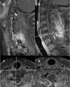
Figure 3. Postoperative MRI after the initial surgery
Postoperative contrasted sagittal and axial MRI after the initial surgery shows a subtotal resection with residual tumor (red arrows) at the facet and near the left vertebral artery MRI: magnetic resonance imaging
Figure 4
Figure 4. Pathologic staining of the tumor
(A) H&E staining shows dense lymphohistiocytic proliferation with no atypia or mitotic figures identified. (B) CD68 immunostaining reveals diffuse positivity reflecting a mononuclear infiltrate characteristic of giant cell tumors. (C) H&E staining shows multinucleated giant cells (red arrow) along with pigment-laden macrophages (black arrow) H&E: hematoxylin and eosin
Figure 5
Figure 5. Postoperative MRI after the second surgery
Postoperative contrasted MRI sagittal and axial after the second operation demonstrating gross total resection of the tumor MRI: magnetic resonance imaging

References

    1. Pigmented villonodular synovitis, bursitis and tenosynovitis. Jaffe HL. https://ci.nii.ac.jp/naid/10024754960/#cit Arch Pathol. 1941;31:731–765.
    1. Pigmented villonodular synovitis and giant cell tumors of the tendon sheath: radiologic and pathologic features. Llauger J, Palmer J, Rosón N, Cremades R, Bagué S. AJR Am J Roentgenol. 1999;172:1087–1091. - PubMed
    1. Pigmented villonodular synovitis of the spine: a clinical, radiological, and morphological study of 12 cases. Giannini C, Scheithauer BW, Wenger DE, Unni KK. J Neurosurg. 1996;84:592–597. - PubMed
    1. C1-C2 pigmented villonodular synovitis and clear cell carcinoma: unexpected presentation of a rare disease and a review of the literature. Lavrador JP, Oliveira E, Gil N, Francisco AF, Livraghi S. Eur Spine J. 2015;24:0. - PubMed
    1. Pigmented villonodular synovitis originating from the lumbar facet joint: a case report. Oe K, Sasai K, Yoshida Y, Ohnari H, Iida H, Sakaida N, Uemura Y. Eur Spine J. 2007;16:301–305. - PMC - PubMed

Publication types

LinkOut - more resources